The basics of x-rays
Published May 2016
•
Copyright © IOP Publishing Ltd 2015
Pages 1-1 to 1-5
You need an eReader or compatible software to experience the benefits of the ePub3 file format.
Download complete PDF book, the ePub book or the Kindle book
Permissions
Abstract
On 28 December 1895, Wilhelm C Röntgen presented his paper 'Über eine neue Art von Strahlen' ('On a New Kind of Rays') to the Würzburg Physical Medical Society. These rays had been labelled with the letter 'x' to represent their unknown nature, which is understandable at a time when there was no knowledge of this strange radiation, and the nature and characteristics of radiation in general were not understood. The knowledge we have gained about x-rays belies the name, as we now possess extensive information about all aspects of x-rays.
1.1. What are x-rays?
On 28 December 1895, Wilhelm C Röntgen presented his paper 'Über eine neue Art von Strahlen' ('On a New Kind of Rays') to the Würzburg Physical Medical Society [1]. These rays had been labelled with the letter 'x' to represent their unknown nature, which is understandable at a time when there was no knowledge of this strange radiation, and the nature and characteristics of radiation in general were not understood. The knowledge we have gained about x-rays belies the name, as we now possess extensive information about all aspects of x-rays.
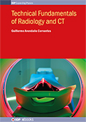
X-rays are a form of radiation or electromagnetic wave, as are radio waves and visible light. Electromagnetic waves are variations in the amplitude of energy in time and are classified according to the speed with which they fluctuate over time. This defines the concept of the wavelength, the time taken by a wave to complete a full sequence (see figure 1.1). As a wave oscillates faster its wavelength becomes smaller and its frequency increases. More specifically, all electromagnetic waves lie in the electromagnetic spectrum, which is arranged by the wavelength or its equivalent, the frequency. X-rays lie above ultraviolet radiation, visible light and radio waves in the spectrum, and below cosmic radiation; they are a form of high energy radiation, with a high frequency and short wavelength.
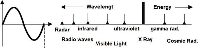
Figure 1.1. An electromagnetic wave and the electromagnetic spectrum.
Download figure:
Standard image High-resolution imageWe now know that the best way to produce x-rays is by accelerating electrons, which produce the desired radiation on impact with a target made of a suitable substance. This process is performed inside a vacuum tube in which two electrodes are installed. A high electric potential is applied to the electrodes to accelerate electrons from the cathode which then impact on the anode, producing x-rays. All this is possible if there is a system outside the tube controlling the process. This is similar to the conditions inside the tube of an old-fashioned TV screen, the cathode ray tube (the generation of electrons, the application of a high voltage, and the screen as the target), in which small amounts of x-rays are produced. These can be a cumulative danger to health, which is why parents are advised not to allow children to get too close to the screen when watching TV.
The exact way to produce a given quantity and quality of x-rays will be discussed later, when analyzing the x-ray tube and generators in detail. When the electrons impact the target—the anode—x-rays are produced in two main ways (figure 1.2):
- (a)Bremsstrahlung. This form of radiation, 'braking radiation' in German, is explained by the fact that the accelerated electrons, as they hit the anode, are slowed down if they have high energy and the amount of braking (energy) is converted into different forms of radiation. This radiation is diverse and of different wavelengths because it originates from different amounts of braking.
- (b)Characteristic radiation. This form of energy is produced by the eviction of orbital electrons through impacts with other electrons sent from the cathode, and a characteristic x-ray emission occurs for specific electron orbits.

Figure 1.2. The mechanisms of bremsstrahlung and characteristic radiation.
Download figure:
Standard image High-resolution imageFor medical diagnosis, the x-rays typically used are composed of 70% bremsstrahlung and 30% characteristic radiation [2].
1.2. Characteristics and properties of x-rays
- –X-rays are electrically neutral, that is, they do not experience deviation or deflection when inside an electric, magnetic, or combined field.
- –X-rays travel in straight lines at the speed of light, a characteristic which can be used to direct and focus the rays in order to radiate the specific region of the body being studied.
- –X-rays produce biological and chemical effects, which means they can affect an organism by producing ionization and/or cellular changes that may be responsible for disorders or further mutations.
- –X-rays span a section of the electromagnetic spectrum and possess not only one frequency, but several. These depend on the set of factors that led to the generation of radiation. As shown in figure 1.3, the higher the voltage that produces the rays, the shorter the wavelength is, i.e. the frequency is higher.
- –X-rays are not visible to the human eye or to animals [3], so their detection is possible only by means of instruments and photographic methods. This is an important consideration in undertaking protective measures for the human body.
- –X-rays produce images on photographic film and fluorescence on certain types of crystals; both phenomena are used as a means to obtain x-rays films and fluoroscopy images on medical monitors, respectively.
- –X-rays produce secondary radiation and scattered radiation, which means that a biological object receiving x-rays produces, in turn, new rays with different characteristics [4]. These rays are generally a problem in imaging and are detrimental to the safety of people who work with x-rays.
Figure 1.3. The relationship between wavelength and applied voltage.
Download figure:
Standard image High-resolution image1.3. Substances and x-rays
X-rays propagate in a straight line and if any substance lies in their path they will behave according to the characteristics of the substance. In this way they can produce some of the effects already mentioned, such as fluorescence or triggering secondary radiation. Three possible interactions may occur: x-ray absorption, transmission, or dispersion. Radiology takes advantage of these phenomena to produce images, either temporary or permanent, which have diagnostic value.
X-ray emission is obtained mainly by bombardment with high-speed electrons on targets [5] made of materials with a high atomic number. As shown in figure 1.4, the x-rays can pass through the substance without being absorbed, in which case we have transmission. If the rays do not penetrate and are stopped by the substance, we have absorption. Dispersion occurs when the x-rays pass through the substance producing secondary and scattered x-rays. The nature of the secondary x-rays depends on their energy and wavelength, as well as the substance the primary x-rays found in their way. Both types of x-rays, secondary and scattered, deviate geometrically from the initial focus of the primary x-rays, which is of great practical importance in obtaining radiographs.

Figure 1.4. X-ray interactions with substances.
Download figure:
Standard image High-resolution image1.4. The effect of x-rays on radiographic film
A fundamental use of radiation is in radiography. It is necessary to determine the penetration of x-rays in living tissue, so we can understand how both the properties of the x-rays and the characteristics of the different tissues produce a combination of effects that determines the degree of blackening of radiographic film. Simply put, the further the x-rays penetrate, the more blackening is produced. In contrast, if the radiation is absorbed by the tissue and barely reaches the film, it produces a white image. Obviously, different shades of gray are the result of intermediate values of attenuation of radiation on tissue [6]. There are basically three factors that determine the degree of blackening of the film:
- (a)The physicochemical properties of the target tissue (atomic number, molecular density, etc), which determine the absorption degree of the radiation.
- (b)The energy or wavelength of the x-rays, which determines whether they will penetrate more or less into the tissue.
- (c)Scattered radiation impinging on the film (in addition to the main radiation) causes the deterioration of radiographic quality.
The above description is best understood by studying any radiographic film. We can see how bones are displayed in white, as little radiation passes through them, while the soft tissues are black, as much more radiation passes through them and reaches the radiographic film [7]. Thus bone is described as a radiopaque tissue, soft tissues and gases are radiolucent tissues, and other tissues that provide images of different shades of gray are defined as radio-intermediate.
Regarding the above factors involved in blackening in typical practice, factor (a) cannot be altered, but factor (b) depends largely on the values selected by the technician, the voltage values and filament current [8]. Factor (c) is neutralized by various techniques which will be discussed later.
Radiation with a short wavelength and high energy has high penetrability [9], so that blackening of the film will occur if the tissue the x-rays go through is low density, producing a grayscale decreasing as the density increases, until the increase in density is such that the radiation does not pass through, resulting in white on the film. Radiation of low wavelength produces, with the same pattern of increasing densities, a grayscale that is less pronounced than in the previous case, with high densities of white and black, this is shown in figure 1.5.

Figure 1.5. Penetration of shorter and longer wavelength x-rays and the effect on the grayscale.
Download figure:
Standard image High-resolution image
References
- [1]Jensen F 1995 100 years of x-rays Medicamundi 40 156–70
- [2]Lidio G, Lidio Y and Mosca E 2001 Técnica Radiológica (Buenos Aires: López Librero Editores)
- [3]Mulkay J and Fernández J 1999 Rayos X (Havana: Pueblo y Educación)
- [4]Tremolieres J 1982 Electrónica y Medicina vol 11 (Madrid: Paraninfo)
- [5]Barroso E 1999 Radiología de la Silla Turca (Havana: Científico Técnica)
- [6]Christensen E and Curry T 1998 An Introduction to the Physics of Diagnostic Radiology (Philadelphia, PA: Lea & Febiger)
- [7]Vegh A 1977 Physical principles of x-ray image formation Medicor News 2 47–52
- [8]Degenhardt H 1983 El desarrollo de las sustancias luminiscentes y la utilización de éstas en la técnica de rayos X Electromédica 4 155–8
- [9]Bushong S C 2008 Radiologic Science for Technologists: Physics, Biology, and Protection (Maryland Heights, MO: Mosby)