
Abstract
A simple photoacoustic method was used to evaluate bovine cortical bone samples. This study aimed to evaluate the effects of glycation in bones due to non-physiological crosslinks on the properties of collagen. The average amplitude of the ultrasonic waves generated by the near-infrared pulsed laser irradiation was smaller in the glycated bone sample than in the reference (normal) sample. The results indicate that glycation due to diabetes might affect the photoacoustic properties of the bone. Ultrasonic waves with a small amplitude were also generated in the bone because the bone sample was not perfectly opaque to the light used.
Export citation and abstract BibTeX RIS
Diabetes causes various complications, such as retinopathy, nephropathy, and neuropathy. In particular, the accumulation of advanced glycation end products (AGEs) crosslinks in collagen 1) constitutes a major problem; typical AGEs crosslinks include pentosidine and glucosepane. 2) Glycation due to AGEs crosslinks is generated by aging and renal failure. In contrast to physiological crosslinks, AGEs crosslinks change the viscoelasticity of collagen in the body. Bones are mainly composed of minerals such as hydroxyapatite and type I collagen. 3) The National Institutes of Health reported the dependence of bone strength on bone mineral density (BMD) and bone quality. 4) Bone quality is affected by the material properties and microscopic and macroscopic structures 5–8) in the bone; it is also affected by remodeling, cell function, and the environment surrounding cells and bone matrices, such as oxidative and glycation stress. Therefore, diabetes accompanied by glycation may affect bone quality. Wen 9) and Atsumi 10) reported an increase in bone fracture risk in the diabetic patients in their study, even though the patients had normal BMDs, implying that bone strength cannot be measured accurately using X-ray techniques as these techniques can only evaluate the minerals in the bone. Therefore, studies on collagen properties in bone are being considered for bone quality assessment.
Previous studies have focused on investigating the changes in bone strength and elasticity due to glycation in collagen. 11,12) Recently, the longitudinal wave velocities of ultrasonic waves in glycated bones have been studied. Since the ultrasonic wave properties in the bone are strongly affected by its complicated structure, the evaluation of bone material properties using conventional ultrasonic techniques in the MHz range is difficult. Therefore, both Imoto and Yasui used a micro-Brillouin scattering technique in their studies to obtain the wave properties in minute areas of bone samples. 13,14) Imoto reported a decrease in the longitudinal wave velocity in artificially glycated bovine bone samples; 13) Yasui et al. reported a decrease in the longitudinal wave velocity in the bones of spontaneously diabetic torii (SDT) rats. 14) As the mass density of the bone samples remains almost the same, 15) these studies concluded that the elasticity of the bone decreased because of glycation.
Additionally, the photoacoustic (PA) method, which uses energy conversion from light to sound in materials, may be applied for physically evaluating collagen properties in bone; it is a non-contact method capable of achieving high-resolution microscopic imaging. The application of this method for bone evaluation is not as popular as it is for evaluating soft tissues; 16,17) however, some applications have shown the possibility of bone microstructure evaluation. 18) Lashkari investigated the effect of collagen in bone on PA properties using pulse lasers with wavelengths of 1064 and 805 nm. 19) Since light with a specific wavelength can pass through the skin, Steinberg et al. reported an in vivo bone evaluation system using the PA technique. 20) However, PA studies on diabetic bones have not yet been conducted.
In a previous report, we investigated the effects of glycation on the PA characteristics of bone. 21) In this work, we investigated light transmission through the bone in addition to analyzing the ultrasonic waves generated in the bone. Wavelengths of light between 1000 and 1700 nm show a deeper light transmission inside the tissue. 22) However, the absorption coefficient of water tends to increase as the wavelength increases. Therefore, we used a light source with a wavelength of 1064 nm. At this wavelength, collagen mainly absorbs light resulting in ultrasound generation.
The cortical bone from the left femur of a 46-month-old bovine was cut with a band saw (A16, Akiyama Machinery, Japan). The bone was then processed into a rectangular shape with an accurate cutter device (Accutom 10, Struers Japan, Japan). Finally, two plate samples (7.0 × 3.5 × 0.5 mm3) were fabricated from a neighboring site in the bone. The normal directions of these plate samples were in the axial direction of bone (body-weight direction).
The two samples were used as the glycated and reference samples. Each sample was incubated in different solutions. The reference sample was placed in a mixture of phosphate-buffered saline (PBS; 166-23555, Wako, Japan) and penicillin-streptomycin (PS; 168-23191, Wako, Japan). The glycated sample was placed in a mixture of PBS, PS, D-(-)-ribose (R9629; Sigma-Aldrich, USA), and Protease Inhibitor Cocktail Set III (539134, Calbiochem, USA). These samples were then kept in an incubator at 37 °C for 14 d. Imoto et al. fabricated glycated and reference cortical bovine bone samples in the same manner and measured the number of collagen crosslinks using high-performance liquid chromatography. 13) They reported that the number of AGEs crosslinks in the glycated sample was about two-fold that of the reference sample. The increasing tendency of the AGEs crosslinks did not depend on the site in the bovine femur.
The light transmittance in the bone was measured using a pulse laser (1064 nm, Cobollt TorTM XS, HÜBNER PHOTONICS, Sweden, pulse width, 1.4 ± 0.3 ns, 58.3 mW) and a power meter (Thorlab, PM100D, USA). We ensured that the light beam passing through each sample was in the direction of thickness.
Figure 1 shows the photoacoustic experimental system used in this study; the same pulse laser was used for ultrasound excitation in both samples. The focused beam diameter at the surface of each bone sample was 80 μm. The samples were set on a stage with a hole in degassed water at 24.6 °C. This water immersion technique was suitable for avoiding the effects of air invasion caused by the presence of several small pores in the Haversian canals. Polyvinylidene difluoride (PVDF) self-made transducers (diameter 3 mm) were placed above and under the bone samples to record the waves generated at the top surface of each sample and the waves that pass through it. Nakamura reported flat frequency responses of PVDF transducers. 23) The distances between each sample and the transducers were 4.2 mm (above bone) and 4.5 mm (under bone). The PVDF transducers were set near each sample in water to observe weak photoacoustic signals generated in the bone. After amplifying the observed waves using a pre-amplifier (SA430F5, NF Co., Japan), the waveforms were observed using an oscilloscope (DPO7254C, Tektronix Co., USA). By changing the measurement position on the bone sample, generated waves at 20 different positions were observed for each sample.
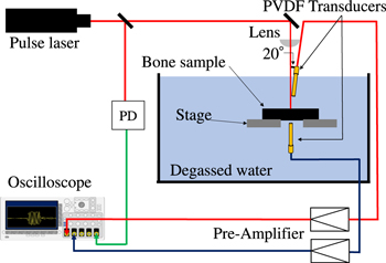
Fig. 1. (Color online) Experimental setup for ultrasound generation and measurements; two transducers were set above and under the sample.
Download figure:
Standard image High-resolution imageFirst, we experimentally observed the transmission of light through the samples. The observed values of the transmitted light power in the thickness direction were weak: 1.63 mW with a standard deviation (SD) of 0.09 mW for the reference sample and 1.34 mW with an SD of 0.04 mW for the glycated sample; as mentioned above, the thickness of each sample was 0.5 mm. These results indicate that the bone samples were not perfectly opaque and that the ultrasonic waves might have been generated at the surface and inside the sample. In addition, the transmitted light intensity of the reference sample was higher than that of the glycated sample, suggesting that glycation might have affected the optical properties of the bones. However, the transmitted light includes the effects of reflectance, scattering, and absorption of light in the bone sample; thus, it is difficult to determine the exact changes occurring in the material properties because of glycation. Some absorption coefficients of healthy human bone, between 0.19 and 0.22 cm−1, have been reported. 24,25) However, the absorption coefficient of glycated bone has not yet been reported.
Figure 2 shows the ultrasonic waves observed above and below the samples. In all cases, the initial waves at 0 μs (time of laser irradiation) were followed by weak low-frequency negative waves. The initial wave appears to be the electric noise due to laser irradiation. The low-frequency negative waves are long, possibly resulting from a different mechanism caused by the laser noise. We tried to observe similar signals using a PVDF film (KF piezofilm, Kureha, Japan, thickness 40 μm).
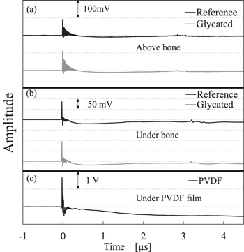
Fig. 2. Observed waveforms (a) above the bone samples, (b) under the bone samples, and (c) under the PVDF film.
Download figure:
Standard image High-resolution imageIt is evident from Fig. 2 (c) that the PVDF film showed a strong low-frequency negative wave after the initial wave. Considering the slow changes, the negative signals were possibly due to pyroelectricity. Lang reported a pyroelectric coefficient of a human femur of 0.0036 μC/m2K, 26) whereas Bravina reported a PVDF pyroelectric coefficient of 30 mC/m2K. 27) Since the thicknesses of the sample and PVDF film were different, it was difficult to describe the phenomena quantitatively. However, the strong negative component observed in the case of the PVDF film can be explained by its strong pyroelectricity.
Considering the distance between the bone sample and PVDF transducers for ultrasound measurements, small waves appearing around 2.8–3.5 μs in Fig. 2 seem to be ultrasonic waves generated by the laser irradiation (photoacoustic waves). Figure 3 shows the detailed features of the waves; here, the low-frequency components were removed by fitting. The waves observed above the bone include the initial largest wave, smaller next waves, and large waves followed by the small waves. The largest wave around 2.8–2.9 μs was generated at the surface of the bone, considering the distance between the transducer and the laser irradiation point on the bone. The smaller waves around 2.9–3.0 μs might have been generated inside the bone because the large wave around 3.0–3.1 μs may be the longitudinal waves generated at the bone surface and reflected at the bottom surface, considering the longitudinal wave velocity of the bone (3800–4400 m s−1). 28) Finally, the waves around 3.2 μs seem to be the shear waves generated at the top surface of the bone, considering the shear wave velocity (1800–2000 m s−1) 29,30) and the propagation time.

Fig. 3. (Color online) Observed waveforms (a) above the bone samples and (b) under the bone samples.
Download figure:
Standard image High-resolution image
Under the bone, small waves were observed around 3.1–3.4 μs; these waves were mostly generated at the top surface of the bone sample. Although the observed waves changed because of the different irradiated positions on the top surface, the observed waves of the reference bone sample were always larger than those of the glycated bone sample.
Next, we compared the maximum amplitudes of the generated waves. Here, the amplitudes were normalized by the average value of the maximum wave amplitudes observed above the reference sample. Above the glycated sample, the normalized maximum amplitude of the observed wave was 0.74 with an SD of 0.14, indicating that changes in the photoacoustic properties of the bone occurred because of glycation. As mentioned above, glycation causes the degradation of collagen by AGEs formation. The present results indicate a possibility of performing a quality evaluation of collagen in bone by the generated wave. As near-infrared light passes through the skin to the bone, 20) a simple combination of a near-infrared pulse laser and ultrasonic transducer may enable the evaluation of bone glycation in vivo.
The normalized maximum average amplitude of waves observed under the reference sample was 0.24 with an SD of 0.05, while that of the waves observed under the glycated sample was 0.14 with an SD of 0.02. Thus, the waveforms observed under the bone showed a decrease in amplitude. The maximum amplitudes resulted from the wave generated at the top surface of the bone sample; subsequently, the decrease resulted from attenuation in the bone. The amplitude ratios of the glycated and reference samples were 0.74 (observation above the samples) and 0.58 (observation under the sample). These results indicate that the wave attenuation might have been stronger in the glycated sample than in the reference sample. From high-frequency Brillouin scattering measurements, Yasui reported that glycation reduced the longitudinal wave velocity in young SDT rat bones, indicating the possibility of "softening" in the glycated young bone. 14) Our present results were obtained from an artificially glycated bovine bone; however, the stronger wave attenuation might have resulted from the "softening" reported by Yasui as it is consistent with his data.
According to our research, this is the first experimental photoacoustic study of a glycated bovine cortical bone. Although only one reference sample and one glycated femur sample were used, the measurements were performed at different positions in these samples, considering bone heterogeneity. The results indicate that the photoacoustic technique using near-infrared light may be a good tool for the in vivo evaluation of diabetic effects on long bones such as the tibia because light can pass through the thin, soft tissues of the bone. Future studies could research treating several bone samples from various sites and attempting an in vivo evaluation of the bone.
Acknowledgments
This study was partly supported by Heiwa Nakajima Foundation.