Abstract
We have heard it over and over again – internal combustion engines (ICE) operated with fossil fuels can cause severe damage to our health and environment.1 Nevertheless, they are still very widespread and will not disappear until a compelling alternative is found. One possible alternative is the low temperature polymer electrolyte fuel cell (LT-PEFC). They are commonly operated with hydrogen and air and only emit water. However, before LT-PEFCs can replace ICEs on a large scale mainly two barriers need to be overcome. Those barriers are cost and lifetime. Hence, reducing the costs of a fuel cell and increasing its lifetime are the two most relevant research fields regarding fuel cell commercialization. Whereas the costs are mainly dictated by the raw material prices (e.g. platinum) and the manufacturing process the reasons for limited lifetime are numerous.
PEFCs in general are affected by various degradation effects at different locations of the fuel cell.2,3 The membrane suffers for example from pinhole formation and membrane thinning. The hydrophobicity of the gas diffusion layer decreases over operation time which decreases its water transport ability. The bipolar plates are prone to surface corrosion which reduces their conductivity. The catalyst and catalyst layer also represent a major degradation area. Some specific degradation mechanisms are carbon corrosion, catalyst detachment and catalyst particle growth. All degradation effects are complex functions of the specific operational parameters, making it challenging to identify them individually. This in turn is necessary to gain insights into limitations in fuel cell lifetime and is the key to successful development of mitigation strategies to reach a desired lifetime of >5,000 hours for automotive drive cycle operation.
There are different techniques available to identify and characterize the individual degradation mechanisms. Some common ones are polarization curve or electrochemical active surface area measurements which are based on electrochemistry. Furthermore, imaging techniques like secondary electron microscopy (SEM) are frequently used for degradation analysis. Another established imaging technique is X-ray tomography. The main advantages of X-ray tomography over SEM imaging are its ability to capture the fuel cell area of interest as a whole and that the tomography scan can be recorded nondestructively and in-operando.4
For some time, fuel cell X-ray tomography was mostly carried out at large scale synchrotron beamline facilities. Unfortunately, they tend to have only very limited access and therefore only few experiments can be conducted in a short time period. In recent development though, lab sized X-ray microscopes have gained interest in the community.5 Such microscopes have excellent availability and therefore enable the possibility to carry out long term experiments.
The present contribution will utilize this particular advantage. Our unique approach is to retrace fuel cell degradation with X-ray tomography imaging (cf. Figure 1) over time and correlate it with individual failure mechanisms. In particular, electrode degradation will be discussed. MEAs, with e.g. different carbon/platinum ratios, were exposed to accelerated stress tests and imaged at beginning and end of life. With the aid of X-ray tomography, we are able to segment the MEA and subsequently locate and analyze the degradation effects at the electrode. In combination with electrochemical measurements, exclusive in depth insights of fuel cell degradation are provided. In addition, a novel approach for post X-ray tomography processing is discussed. In summary, the combined findings lead to an improved understanding of LT-PEFC degradation which is a necessary first step in order to investigate new mitigation strategies to increase the fuel cell lifetime.
Figure 1. A membrane electrode assembly visualized by X-ray computed tomography recorded at beginning of life.
1. S. M. Platt et al., 'Two-stroke scooters are a dominant source of air pollution in many cities', Nat. Commun., 5, p. 3749, 2014.
2. J. Wu et al., 'A review of PEM fuel cell durability: Degradation mechanisms and mitigation strategies', J. Power Sources, 184, pp. 104–119, 2008.
3. T. Engl, L. Gubler, and T. J. Schmidt, 'Think Different! Carbon Corrosion Mitigation Strategy in High Temperature PEFC: A Rapid Aging Study', J. Electrochem. Soc., 162, pp. F291–F297, 2015.
4. J. Eller et al., 'Progress in In Situ X-Ray Tomographic Microscopy of Liquid Water in Gas Diffusion Layers of PEFC, J. Electrochem. Soc., 158, B963 (2011).
5. M. Andisheh-Tadbir, F. P. Orfino, and E. Kjeang, 'Three-dimensional phase segregation of micro-porous layers for fuel cells by nano-scale X-ray computed tomography', J. Power Sources, 310, pp. 61–69 (2016)
Funding for this research was provided by the Natural Sciences and Engineering Research Council of Canada, Canada Foundation for Innovation, British Columbia Knowledge Development Fund, and Ballard Power Systems through an Automotive Partnership Canada grant.
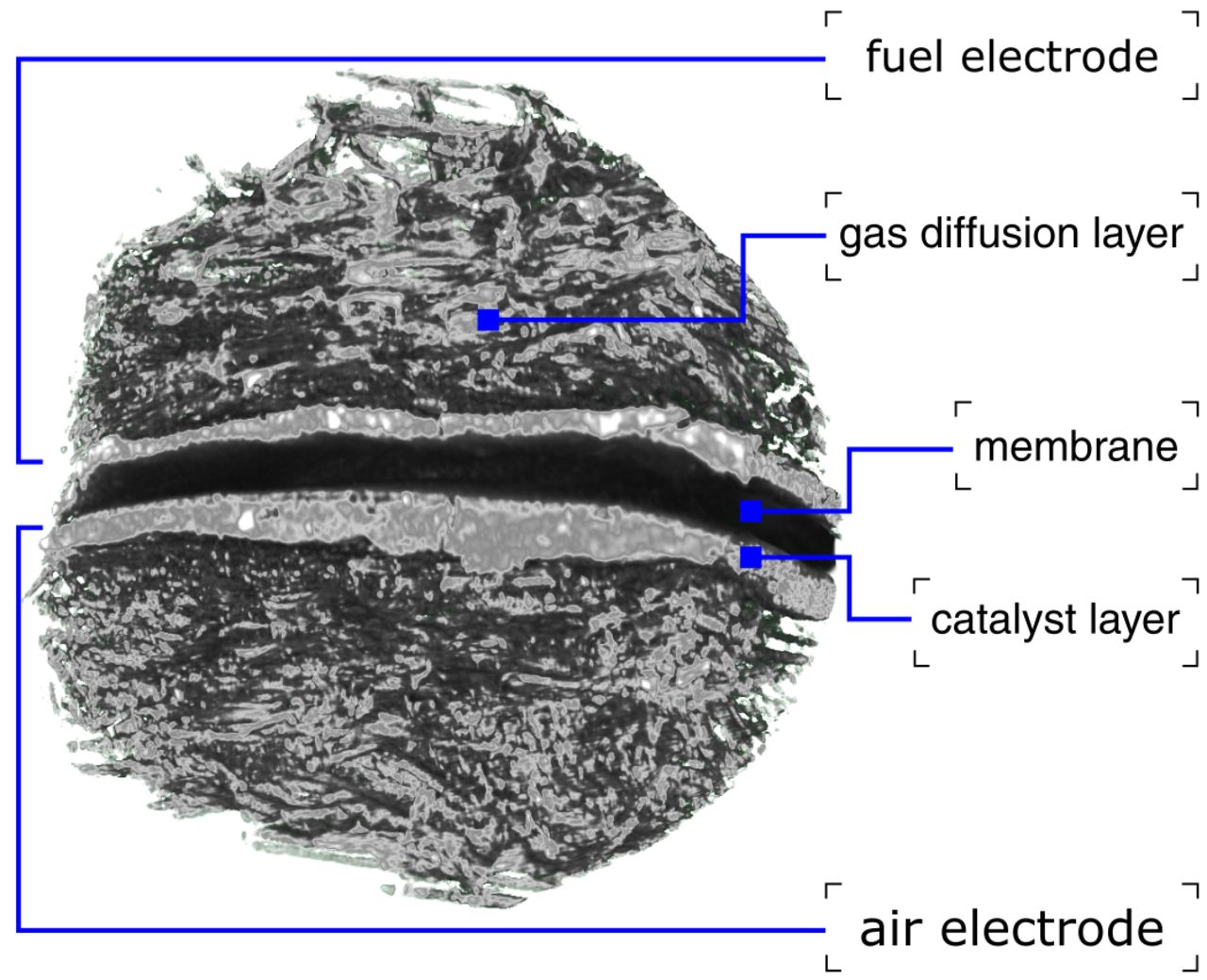
Figure 1