
Abstract
Morphine is a powerful opioid pain medication and commonly used narcotic pain killer and is toxic during overdose or when abused. Compared to conventional analytical techniques, the electroanalytical method has significant advantages viz. low cost, simplicity, ease of operation and facile miniaturization. In the present paper different approaches based on various modifications adopted for effective electrochemical sensing of morphine are reviewed in a comprehensive way. Among different modified electrodes available for the detection of morphine, carbon based materials—CNTs and graphene—display effective quantification and are attractive in terms of cost compared to noble metals. In addition, the performance of reported sensors in terms of their including detection range (LDR), limit of detection (LOD) and technique used are presented. The present review compares various electroanalytical techniques adopted for the determination of morphine.
Export citation and abstract BibTeX RIS

This is an open access article distributed under the terms of the Creative Commons Attribution 4.0 License (CC BY, http://creativecommons.org/licenses/by/4.0/), which permits unrestricted reuse of the work in any medium, provided the original work is properly cited.
Morphine, an alkaloid derived from the poppy plant is the world's first true and real drug. Morphine is a naturally occurring phenanthrene derivative (Fig. 1a). It has been available for centuries and appeared in Pliny's Historia Naturalis (AD 77) as opium, the resin derived from poppy sap. The Friedrich Wilhelm Adam Sertürner, a German pharmacist and a pioneer of alkaloid chemistry took the first step in identifying opium's active ingredient, morphine in 1803.1 It is currently obtained using techniques greatly unchanged over eight millennia.2–5

Figure 1. (a) The lanced pod of Papaversomniferumshowing extrusion of the opium containing resin. By KGM007 [Public domain], via Wikimedia Commons. (b). Structure of Morphine.
Download figure:
Standard image High-resolution imageThe extraction of morphine was first achieved by Nicolas Lemery (1645–1725) due to the differential solubility of morphine in water, alcohol, and organic solvents.2 In Europe, the commercial production of morphine began as early as in1820s and the 1830 s in the different parts of the world.6–10 The combination of the greater potency of morphine over opium and the ability to inject the solution directly in to the blood stream provided swifter pain relief.6 In 1912 the United States and many other countries signed the International Opium Convention which controlled the import, manufacture and sale of morphine.11 The World Health Organizations recommended morphine as the most commonly used opioid for moderate to severe pain in people with cancer in the UK and other European countries and in the United States for non-malignant pain.11
Morphine acts directly at pain modulating receptors in the nervous system, termed opioid receptors.12–14 These receptors are distributed throughout the central nervous system (CNS) with high concentration in the nuclei of tractussolitarius, peri-aqueductal gray area (PAG), cerebral cortex, thalamus and the substantia gelatinosa (SG) of the spinal cord. They have also been found on peripheral afferent nerve terminals and many other organs. When morphine binds the opioid receptors, the pain killing message is transmitted inside the cell through G protein cascade. The G protein is the most common method of signaling in our cells. It increases conduction through potassium channels, decreases conduction through calcium channels, and inhibits adenylyl cyclase. These changes blunt the effect of signaling systems that transmit pain.13,14
Morphine is additive and leads to drugs abuse. When morphine acts in the reward center of the brain-the area that makes eating and other essential processes feel pleasurable. The brain reacts to morphine by building more components for the G protein signaling system. As time goes on more and more morphine is needed to have the same effect on the system. When morphine is removed, the normal function of the pleasure system is blunted by the bloated G protein signaling system leading to severe withdrawal systems.
Morphine usually as the sulfate or hydrochloride can be given orally, intramuscularly, intravenously, subcutaneously, rectally, epidurally and intrathecally. The intramuscular dose is 0.1 to 0.2 mg Kg−1, peak effect is in 30–60 min and duration action is 3–4 h. In intravenous administration total dose is similar. Morphine can be given epidurally at 10% and intrathecally at 1% of the parenteral dose. The dose to be used is determined by the patient's age, not their weight. The dose is adjusted according to the response, so patients need to be observed more frequently than four-hourly.15
In clinical medicine morphine is regarded as the gold standard or bench mark of analgesic used to relieve severe or agonizing in cancer patients. It is used as the starting compound for the synthesis of other opioids such as hydromorphine, oxymorphine and heroin, whose analgesic effect and addiction are so strong so as to be considered as drugs of abuse.16–19 On the other hand morphine is toxic when overdose or abused and can also influence various immune functions as well as disruption in central nervous system, respiratory rate and low blood pressure. In addition morphine analysis in biological fluids has been applied both in forensic cases and pharmokinetic studies. All these facts points towards the need for developing a sensitive, selective and convenient electrochemical sensor for the strict monitoring of morphine in many environmental and biological samples is necessary.
Electrochemical Methods—a Better Approach for Detecting Morphine
Several analytical methods like chemiluminescense,20 surface plasmon resonance,21 immuochromatography assay22 gas chromatography,23 HPLC with UV,24 radioimmunoassay,25,26 HPLC with amperometry,27 resonance light scattering,28 liquid chromatography coupled with UV detection29 thin layer chromatography30 resonance scattering,31 time resolved chemiluminescence,32 spectrometry,33 immunoassay34 are being employed for the detection of morphine. Although these methods can offer good selectivity, they often require advanced technical expertize, complex pretreatment steps, time consuming, and are expensive. Thus an alternative strategy for the detection of morphine is necessary. Unlike surface plasma resonance, chromatographic techniques, electrochemical sensors can be employed as an alternative approach due to their high selectivity, low cost and fast response for detecting a wide range of analytes. Electrochemical sensors have become remarkable in fields of medicine and biotechnology, environmental monitoring and for industrial applications. The possibility of electrode fabrication by using competent modifications help to tailor the properties of working electrode and explore its novel electrocatalytic effects towards sensing of analytes. In addition these sensors are capable of being incorporated into stable, portable or miniature devices enabling specific applications. Even though many reports are available for the electrochemical detection of morphine a comprehensive review deals with different sensors for morphine is not available. The two reviews now available for reference review morphine sensors to some extent along with other neurological drugs and neurotransmitters35 whereas the other deals with immunosensors.36 A comprehensive review exclusively devoted for morphine sensors will be beneficial in the present scenario. The present paper therefore reviews the progress in electrochemical detection of morphine emphasizing the different modified electrodes.
Electrochemistry of Morphine
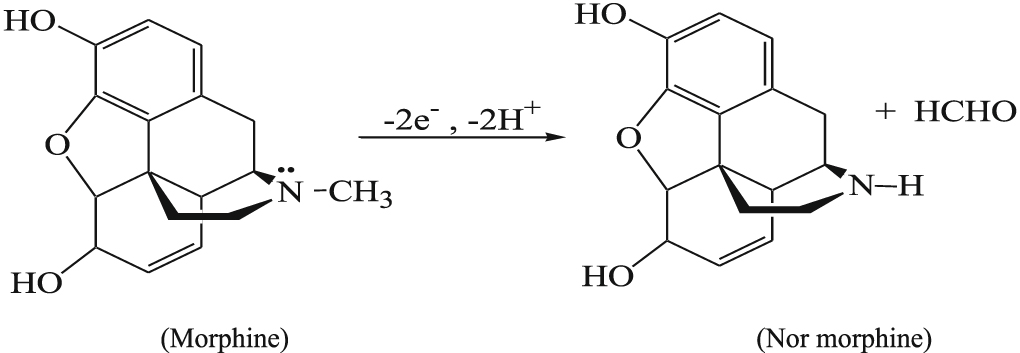
Morphine being electroactive in which both phenolic and tertiary amine group present are responsible for its catalytic activity. The oxidative mechanism proposed involves one proton and one electron corresponding to the oxidation of the phenol ring followed by dimerization of the free radical to pseudomorphine. Whereas two proton, two electron reaction takes place during the oxidation of the aliphatic tertiary amine group in morphine and gives rise to normorphine as the oxidative product as illustrated. (Schemes

Scheme 1. Mechanism of oxidation of the phenol ring of MO.
Download figure:
Standard image High-resolution image
Scheme 2. Mechanism for the oxidation of the tertiary amine group of MO.
Download figure:
Standard image High-resolution imageMorphine (MO) usually co exists with its common interferents such as diclofenac (DF), ascorbic acid (AA), uric acid (UA), glucose (GLU), salicylic acid (SAL) they undergo similar two proton two electron reaction at most of the electrodes. Therefore a selective and sensitive method which is capable of detecting morphine in presence of its interferences is more advantageous. Electroanalytical methods serve as better and excellent methods to meet this crucial need and are more suitable for the detection of the electroactive molecules.
Cyclic voltammery (CV) is the most important and widely used electroanalytical technique that gives first hand information about the oxidation process. Though CV is highly informative, its high back ground current imposes some limitations for its use in the determination of various molecules. Therefore more sensitive technique like differential pulse voltammetry (DPV), square wave voltammetry (SWV) and chronoamperometry can be employed for the low level detection of a wide range of analytes.
The working electrode plays a major role in the detection process and has enormous scope for modification by materials at the nanoscale. The modified working electrode enables the resolution of the signals of each analytes whereas only single overlapped signals are obtained at solid electrodes.
Diversely Modified Electrodes for MO Determination
The role of modified electrode is very significant as far as a sensor is concerned. The improvement of quality of life has stimulated a large quantum of research in this area. The properties desirable for a particular analyte can be attained by a suitable modification. The emergence of nanoparticles and subsequent developments in the field of material science has contributed tremendously to the present scenario of sensors. As working electrode is the most important part of a sensor many modifications enabled selective and sensitive determination of morphine. Common working electrodes range from inert metals such as gold, silver or platinum to diverse forms of carbon materials.
Solid Metallic Electrodes Used for the Electrochemical Determination of Morphine
The most commonly used metallic solid electrodes are gold, platinum and aluminum. These metals can be obtained with high purity, machined easily and developed readily into a wide variety of configurations–wires, rods, flat sheets and woven gauzes. So gold, platinum and aluminum electrodes have been modified and used in this phase.
Ganjali et al. developed a sensitive and fast method for the determination of methyl morphine in human urine and plasma through a new electrochemical method based on flow injection analysis (FIA) and fast Fourier transform cyclic voltammetry (FFTCV). The ultra micro electrode (UMEs) involving metal microwires were used. In this method the current changes with various processes occurring on the electrode surface depending on oxidation / reduction of adsorbed analyte and inhibition of oxidation and reduction of electrode surface by adsorbed analyte etc can be assessed. By choosing high scan rate greater than 20 V S−1 can provide information on the influence of adsorbed analyte. Whereas at low scan rate the amount of the desorption analyte molecule can be changed. Thereby part of adsorbed analyte molecule still remain on electrode surface inhibit redox process on the electrode surface. In this method ΔQ is calculated according to the current changes in the cyclic voltommogram. The selectivity and sensitivity of the analyte response expressing in terms of ΔQ strongly depends on the selection of integration limits. One of the important aspects of this method is the application of a special digital filtration which is applied during the measurement. The electrochemical oxidation process of gold electrode surface initiates with the hydroxyl ion desorption which at more positive potential resulting gold electrode formation due to structural arrangement. Besides at a more positive potential, surface oxidation can be initiated by adsorbed water molecule leading to formation of gold oxide.

Use of this detection method demands a high scan rate is the drawback of this method.37 Whereas chemically modified electrodes showed improved catalytic activity with lower potential and much higher current response compared to bare metallic solid electrodes. The enhanced electrocatalytic activity for morphine oxidation at 2-aminoethanethiol or cysteamine self assembled monolayer (SAM) modified gold electrode was studied by Hong et al. The SAM modified Au electrode showed potent electrocatalytic activity towards the electrochemical oxidation of morphine in phosphate buffer solution (pH 6.0) with decrease of the overpotential and two fold increase in the oxidation current compared to the bare gold electrode.38 The SAM blocks access of solvent molecule and electrolyte ions from reaching the surface, thus protecting surface from oxidation. Another added advantage is preventing the electrode from surface fouling which is favorable for analyte oxidation. Thus oxidation of MO is favored in a SAM modified electrode. The surface charge state of 2-amino ethane thiol SAM is pH dependent, lower the pH, the more positively charged surface which is favorable for MO adsorption through hydrogen bonding.
The double stranded DNA immobilized onto the mercapto-benzaldehyde modified gold electrode was successfully used for the sensitive determination of morphine. The sensor was fabricated by immersing gold electrode (Au) in 1 mmol L−1 mercapto-benzaldehyde (MB) for 12 h at room temperature, where the assembly of MB monolyayer was performed via sulfur—Au reaction. Then the obtained electrode as completely cleaned with double distilled water to remove un assembled thiol component and was denoted as (Au/ MB). Then Au/MB electrode was incubated at 10−7 mol l−1 5'- NH2 modified single stranded dNA (ss-dNA) for 12 h and washed with phosphate buffer solution (PBS) and was denoted as Au/MB/S1. This modified electrode was immersed in 0.1 mol l−1 PBS (pH =5) containing 10−7 mol l−1 double stranded dNA (ds-dNA) with shaking for 30 min at 37 °C. The obtained electrode was washed with same PBS solution and double distilled water and resulting electrode was denoted as Au/MB/ds-DNA. The interaction of morphine with DNA onto the electrode surface has been studied by differential pulse voltammetry. This suggested method possesses many advantages such as low detection limit, faster response, low cost and simplicity.39 Among the three above mentioned sensors the last one is attractive in terms of its wide linear range 0.05–500 μM.
Aluminum in conjunction with palladium and Prussian blue was also used for the determination of morphine.40
Prussian blue thin film deposited on ITO glass served as electrocatalytic mediator and an inert optically transparent electrode for the determination of MO.41,42
Metal/Metal Oxide NPs Based Electrode
The incorporation of metal/ metal oxide nanoparticle represents a productive approach towards fabrication of sensitive and selective electrochemical biosensors43–46 Gold nanoparticle, Zinc oxide nanoparticle, platinum nanoparticles, tin oxide, aluminum oxide, nickel oxide, cadmium oxide, titanium platinum/Cobalt alloy nanowire array etc were successfully used for determination of morphine by different researches.16,47–54
The incorporation of metallic oxides to carbon paste has resulted in improved performance of MO sensors. Electrodeposition of gold nanoparticles on carbon paste electrode was another strategy to enhance the sensitivity of an immunosensor with large surface area, good biocompatibility incorporating high conductivity and electrocatalysis characteristics. Atta et al. have explored the feasibility of gold nanoparticles for the determination of MO by incorporating gold nanoparticles in carbon paste electrode.48,49,55,56 Electrochemically deposited gold nanoparticle on a CPE resulted in a sensor with excellent sensitivity for morphine detection where as integration of metalphthalocyanines along with gold nanoparticle (AuMePcMCPE) produced excellent selectivity. The optimized performance of different metal phthalocyanine such as Co, Cu, Ni and Fe at different pH values, the higher current response and lower oxidation potential was obtained by using Co- phalocyanine which is an indication of the best mediation and fastest charge transfer properties (Fig. 2). The difference in the reactivity between different mediator compounds can be attributed to difference in the atomic size of the metals of phtalocyanine compounds which acts as determining factor for the speed of electron transfer processes through carbon paste. This developed sensor possessed a lower detection limit of 5 nM with a linear range of 0.4 μM to 900 μM. Furthermore the sensor can efficiently be used in simultaneous determination of binary mixtures of MO, dopamine (DA), uric acid (UA) and ascorbic acid (AA) with good resolution and peak separation.49 Atta et al. also tried the incorporation of ferrocene on gold nanoparticle in CPE for the determination of MO.56 The current response at bare CPE electrode was 16. 81 μA where as at gold nanoparticle(AuNP) CPE current response increased to 29.03 μA. Furthermore the electrodeposition of gold nanoparticle on ferrocene modified gold nanoparticle resulted in a sharp increase in peak current to 54. 7 μA which indicate an improvement in the electrode kinetics with a substantial decrease in oxidation potential. The apparent diffusion coefficients of MO on CPE increased from 2.96 × 10⁻⁶ Cm2 S−1 to 9.94 × 10⁻⁶ Cm2 S−1 for the AuNp ferrocene modified electrode indicating a quick mass transfer of an analyte molecule towards the surface of the electrode from bulk solution and a fast electron transfer process of electrochemical oxidation of analyte molecule at the interface of the electrode surface and the solution interface. Some researchers also tried gold nanotube arrays for Mo detection.57

Figure 2. (a) Cyclic voltammograms of 1 × 10⁻³ mol l−1 MO in B-R buffer pH 7.4 at scan rate 100 mVs−1 recorded at four different working electrodes: (1) AuFePcMCPE(solid line), (2) AuNiPcMCPE (dotted line) (3) AuCuPcMCPE (dashed line) and (4) AuCoPcMCPE (dashed dotted line). (b) Histogram of 1 mM MO at four different electrodes (AuFePcMCPE, AuNiPcMCPE, AuCuPcMCPE, AuCoPcMCPE) in B-R buffer of pH 2 and pH 7. Reproduced with permission from Ref. 49 Copy right 2014, Wiley.
Download figure:
Standard image High-resolution imageZnO is also one of the most promising material for the fabrication of capable electrochemical sensors due to its special properties.58–61 The electrochemical behavior of morphine at the ZnO doped multiwalled carbon nanotubes modified ionic liquid carbon paste electrode (ZnO/MWCNTs/IL/CPE) was studied using CV and LSV by Barthwal et al. Sol-gel method was used to synthesize the modified electrode (ZnO/MWCNTs/IL/CPE) taking 1-methyl-3-butylimidazolium bromide as an appropriate binder in carbon paste matrix for detection of MO. The MO gives a well defined oxidation peak at the modified electrode due to the oxidation of the phenol ring. The LSV currents of MO at the modified electrode increased linearly with its concentration range of 0.1 to 700 μM and with a detection limit of 0.06 μM. The sensor also provides higher stability, sensitivity, and reproducibility as compared to traditional CPE.47
An electrochemical sensor for the determination of morphine in 0.04 mol l−1 universal buffer solution (pH 7.4) is introduced using gold nanoparticles electrodeposited on a nafion modified carbon paste electrode. The modified electrode shows an oxidation peak due to the oxidation of the phenol ring at the 2-position of morphine, which involves one electron transfer. The oxidation of the phenol ring leads to the formation of pseudomorphine as the main product. At optimum conditions, the concentration of MO was determined using differential pulse voltammetry (DPV) in a linear range of 0.2.0 to 260 μm with a correlation coefficient of 0.999, and a detection limit of 13.3 × 10⁻¹⁰ mol l−1 was obtained. The effect of common interferences on the current response of morphine namely ascorbic acid (AA) and uric acid (UA) is studied. The modified electrode can be used for the determination of MO spiked into urine samples, and excellent recovery results were obtained.55
Magnetic nanoparticles (MNPs) are also tried for improving sensing sensing performance of electrode towards MO. Fe3O4 nanoparticles with low toxicity and biocompatibility was used for enhancing the electrochemical sensing properties towards MO. Dehdashtian et al. have developed a simple and sensitive sensor by incorporating chitosan coated magnetic nanoparticle (CMNP) on CPE for the electrochemical determination of morphine (MO). The electrooxidation of MO was studied on a modified carbon paste electrode using cyclic voltammetry, chronoamperometry and differential pulse voltammetry as diagnostic techniques. The oxidation peak potential of morphine on the CMNP/CPE appeared at 380 mV which was accompanied with smaller overpotential and increase in oxidation peak current compared to that obtained on the bare carbon paste electrode (CPE). Under optimum conditions the sensor provides two linear DPV responses in the range of 10–2000 nM and 2–720 μM for MO with a detection limit of 3 nM. The CMNP/CPE modified electrode successfully measure dopamine (DA) and MO in mixture independently from each other with a potential difference of 200 mV. The proposed sensor was successfully applied for monitoring of MO in serum and urine samples and satisfactory results were obtained.16
NiO is one of the characteristic transition metal oxides that are widely investigated due to its chemical and thermal stability, environmental compatibility and relatively large fundamental band gap (3.6−4 eV). It is successfully used in fuel solar cell, electro-chromic apparatus, magnetic devices, supercapacitor electrodes, lithium ion batteries, gas sensor and heterogeneous catalysis.46,62–65 There are reports on the NiO based electrochemical sensor for the determination of morphine. Sanati et al. have developed an electrochemical sensor based on ionic liquid (IL) 1-methyl-3-butylimidazolium chloride (MBIDZ)Cl modified NiO/CNTs carbon paste electrode (IL/NiO/CNTCPE) for the electrooxidation of MO. The oxidation peak current for morphine increased with increasing amount of NiO/ CNT and Ionic liquid. Also the presence of Ionic liquid/ CPE enhanced the peak current and decreased the overpotential. The presence of NiO/ CNTs on IL/NiO/CNT/CPE surface had great improvement on electrochemical response towards MO. The conductivity of NiO has contributed for very low charge transfer resistance (during impedance measurements) and by addition of IL in to CPE the charge transfer resistance was further reduced resulting in successful oxidation of MO on the surface of IL/NiO/CNT/CPE. Compared to common carbon paste electrodes, the electrochemical response was greatly improved for morphine electrooxidation. The modified electrode successfully resolves the overlapped voltammetric peaks of morphine and diclofenac by 180 mV so that the modified electrode displays high selectivity in the square wave voltammetry (SWV) measurement of morphine and diclofenac in their mixture solutions. The proposed sensor was successfully applied for the determination of morphine in the human urine and pharmaceutical samples.50 A similar study making use of nano nickel oxide single walled carbon nanotubes (NiO-SWCNTs) and 2,4-dimethyl-N/-[1-(2,3-dihydroxy phenyl) methylidene] aniline (DDPM) was done recently by the Akbarian et al. for the determination of morphine, diclofenac and mefenamic acid in pharmaceuticl samples. The three molecule were resolved with sufficient potential separation at ∼247 mV, ∼445 mV and 697 mV at surface of NiO-SWCNTs/DDPM/CPE without any interference. Here NiO-SWCNTs act as a conductive mediator and 2,4-dimethyl-N/-[1-(2,3-dihydroxy phenyl) methylidene] aniline functioned as an electrocatalyst for simultaneous determination of diclofenac, morphine and mefenamic acid.51
Taei et al. followed a facile solid state synthetic route for SnO2-Zn2SnO4 preparation which was further used for modification of multiwalled carbon nanotube carbon paste electrode (MWCNTs/SnO2-Zn2SnO4/CPE) which as subsequently applied for determination of morphine and codeine in human urine samples. The enhanced conductivity offered by SnO2-Zn2SnO4 can be attributed to the presence of donor defects of interstitial tin atoms and the oxygen vacancies. The multiwalled carbon nanotubes effectively increased the electroactive surface area. The synergistic effect of the combination produced a nine times increase in peak current compared to CPE for both the molecules. The nanometer dimensions of MWCNTs, electronic structure and topological defects present on the MWCNTs contributed for the performance of the sensor.66 Similarly morphine and codeine were simultaneously determined by Ali A Ensafi et al. with a modified carbon paste electrode.67 The electrode fabrication involved dispersal of Pt nanoparticles on porous silicon (Psi) by a simple in situ redox reaction between PtCl62− in hydrofluoric acid solution. This Pt/Psi nanocomposite was used as a new electrocatalyst for the determination of morphine and codeine by means of a carbon ionic liquid electrode. This was further integrated in CPE and adsorptive stripping voltammetry was performed. Under optimum conditions, the oxidation current responses of morphine and codeine were linear in the concentration range of 0.10–25.0 μM. The detection limits of 30.0 and 20.0 nM were achieved for morphine and codeine, respectively. The electrochemical sensor has high sensitivity toward the analytes with a good reproducibility (due to the high synergetic activity of Pt nanoparticles and good antifouling properties of the ionic liquid). The proposed sensor was successfully used in real samples containing morphine and codeine.
The voltammetric determination of morphine based on carbon paste electrode modified with ZnO/CNT nanocomposite and 1-methyl-3-butylimidazolium bromide (ZnO/CNTs/IL/CPE) was reported by Afsharmanesh et al. The electrocatalytic activity of MO shows an oxidation peak which is attributed to the oxidation of the phenolic group. SWV results show that the anodic peak current was linear to the MO concentration in the range of 0.1 to 700 μM. The detection limit was calculated as 0.06 μM. The modified electrode (ZnO/CNTs/IL/CPE) was also examined for the determination of morphine in real samples such as urine and ampoules.53
Al2O3 nanoparticle on carbon paste electrode (Al2O3/NP/CPE) was also used for effective determination of morphine by Arabali et al.52 (Fig. 3).

Figure 3. Schematic representation of modified electrode nano metal oxides towards electrooxidation of morphine.
Download figure:
Standard image High-resolution imageAn approach to simultaneously determine paracetamol and MO was initiated by Cheraghi et al. incorporating CdO nanoparticle. They developed CdO nanoparticle-ionic liquid modified carbon paste electrode (CdO/NPs/ILs/CPE) for the voltammetric sensing.68 The results show that CdO/NPs/ILs/CPE could clearly resolve the oxidation peaks of morphine and paracetamol at potentials 0.37 V and 0.65 V respectively. The detection limits for paracetamol and morphine were 0.07 μM and 0.1 μM respectively. The developed sensor was also successfully applied for the determination of the above compounds in tablets and urine samples. The advantage of this modified electrode is the simultaneous determination of morphine in presence of paracetamol with an appreciable peak separation by ∼ 280 mV.
Wester et al. using tetrahedral amorphous carbon (ta-C) which is resistant to wear corrosion and biofouling and possessed the property of supporting facile electron transfer. They report the effect of anodic pretreatment on the electrochemical performance of ultra thin titanium/tetrahedral amorphous carbon (Ti/ta-C) electrodes in terms of surface chemistry and electrochemistry of paracetamol and morphine. The anodic treatment caused a decrease in the oxidation currents of morphine and paracetamol points to the adsorption of paracetamol on carbon is surface chemistry dependent affected by hydrogen bonding, π-stacking and hydrophobic interaction and the sensor offered a very low detection limit of 9.8 nM.69
Carbon Based Electrodes for the Electrochemical Detection of MO
Carbon, an inert electrode material, is used for both oxidation and reduction reactions in both aqueous and non aqueous solutions. It has greater advantages over other metallic solid electrodes due to its unique properties such as high aspect ratio, high strength to weight ratio, extra ordinary mechanical properties, thermal conductivity, optical, magnetic, electrical conductivity, low back ground current, wide anodic potential range, chemical inertness, availability, low cost and relative chemical inertness in most electrolyte solutions. These characteristics have played a major role in the electroanalytical chemistry field, particularly in sensor device. A wide variety of nanoscale materials of carbon with different dimensionalities such as carbon nanotubes, graphene nanosheets etc are available for application as sensors. Carbon based electrodes like spectroscopic grade graphite, pyrolytic graphite, carbon paste electrode, carbon ionic liquid, screen printed electrode, carbon fiber micro electrode, carbon ceramic electrode, and boron doped diamond electrode and glassy carbon electrode are proven to be effective in determining minute quantities of morphine.17,70–74
Pencil graphite leads used as working electrodes are currently known as pencil graphite electrodes (PGEs). Besides the fact that they are cheap, PGEs are also easy to use and more convenient and there is no time-consuming electrode surface cleaning/polishing step. Applying different types of voltammetric techniques in order to quantify a variety of analytes from a wide range of samples, the used PGEs showed reproducible signals, yielding well-defined voltammetric peaks. These electrodes have proven to provide good sensitivity, high electrochemical reactivity, high electrical conductivity, good mechanical rigidity, low cost, low technology, reproducibility, being a viable, renewable and economical tool. The electrochemical characteristics of a PGE, which depends on its surface properties, can be changed/improved either by pretreatment or by chemical modification of the electrode active surface. The PGE has good application in analysis of biomolecules and in the detection of traces of metal ions.75,76
Screen printing or thick film technology is a widely known approach used to manufacture disposable and low cost sensors and biosensors. Replacing conventional electrochemical cells with miniaturized screen printed electrochemical strips is gaining importance in sensor applications in many fields such as pharmaceutical, environmental and food analysis applications. Screen printed electrodes offer a simple user friendly fast and inexpensive route for electrochemical measurement. Ahmar et al. conducted a novel study on determination of morphine using electro membrane extraction (EME) coupled with screen printed electrode. Morphine was extracted from urine sample using this method resulted in a detection limit of 0.005 mg ml−1 with wide linear range of concentrations 0.005 μg ml−1 to 2 μg ml−1. A screen printed carbon electrode was used as working electrode, a carbon counter electrode and silver pseudo electrode was used as reference electrode. Applying EME prior to the electrochemical determination improved the selectivity and sensitivity of the method. Furthermore the sample cleanup ability of EME can increase the life of the electrode and reduce surface passivation. (Fig. 4)70 Another approach to quantify morphine and codeine in urine samples was attempted by Feizbaksh et al. using disposable screen printed carbon electrode strips (SPCE) modified by double strand (ds) calf thymus DNA.71

Figure 4. Schematic illustration of the proposed carrier assisted EME—DPV method. Reproduced with permission from Ref. 71 Copy right 2014, Elsevier.
Download figure:
Standard image High-resolution imageThe practice of using glassy carbon electrode (GCE) has produced many commendable result earlier. The determination of MO using GCE was also attempted by different researches.17,18,72 There was subsequent improvement due to the different modifications on GCE by different researchers. Determination of MO using GCE modified by a pretreatment was attempted by Li et al. Similarly Jordan et al. proposed an electrochemical detection of morphine at a planar glassy carbon electrode. The electrooxidation of MO at GCE involves three steps; first it involves the formation of a phenoxide ion that results from the one-electron oxidation of the phenol ring, followed by the formation of the oxidation product, pseudomorphine, and finally a two-electron loss from a tertiary amine group to normophine as the product. The hydrodynamic voltammogram shows a linear calibration curve in the range of 1.2 × 10⁻¹² to 4.0 × 10⁻¹⁰ moles of morphine injected with a detection limit of 1.24 × 10⁻¹³ moles. The reported method was successfully applied to MO detection in human serum as real sample.18
A comprehensive study of the electrochemical oxidative behavior of morphine in aqueous solution at glassy carbon electrode (GCE) was reported by Garrido et al. The study focuses on the identification of several oxidative products of MO like pseudomorphine, morphine N-oxide, normorphine, dihydromorphine and 2-(N,N-dimethylaminomethyl) morphine, through voltammetric study. The proposed method also verifies that a poorly defined peak observable during morphine oxidation is not a consequence of further oxidation of pseudomorphine but due to the formation of a dimer during the phenolic group oxidation. The results confirm that this method led to the better understanding of its oxidative metabolic pathways.17 A pretreated glassy carbon electrode was used for morphine detection by Li et al.72 The pretreated introduces oxygen containing functional groups on the electrode surface.77 These functional group exhibits certain interaction such as electron donor acceptor, hydrogen bonding, electrostatic dispersive and solvophobic interactions with various species. Morphine could also be effectively adsorbed and accumulated on the electrochemically pretreated glassy carbon electrode. The pretreated GCE could successfully measure morphine and codeine in mixture independently from each other with a potential difference of 530 mV. There are two linear relations between oxidation peak current and MO concentration in the ranges 4 to 18 μM and 18–100 μM with a detection limit of 0.2 μM. This developed method had been applied to the determination of morphine in urine samples.
The performance of the MO sensor improved significantly by adopting mesoporous carbon modified glassy carbon electrode (OMC/GCE) by Li et al. The electrochemical behavior of morphine was studied and a reversible anodic peak was obtained, which was much larger than that on a bare GCE. The voltammetric studies revealed that there were two anodic peaks of morphine at 0.418 V and 0.866 V at OMC/GCE. The presence of two anodic peaks attributes to the oxidation of both phenol ring and aliphatic tertiary amine group which gave pseudomorphine and normorphine as the oxidation products (Fig. 5). These facts clearly indicate that ordered mesoporous carbon has excellent electrocatalytic activity which may be attributed to its large surface area and a large number of edge plane defect sites at its surface. The modified electrode displayed a decrease in the overpotential by 82 mV and an increase in the peak current by 80 times as compared to the GCE. There was a good linear relationship between anodic peak current and morphine concentration in the range of 0.1 μM to 20 μM with a detection limit of 10 nM for both phenol and tertiary amine oxidation peaks. The sensor has also been successfully applied to the selective determination of morphine in urine samples with a low detection limit of 50 nM. The results confirm that the OMC/GCE modified electrode holds a promising candidate in practical application.73

Figure 5. Electrochemical oxidation of morphine at OMC/GCE. Reproduced with permission from Ref. 73 Copy right 2010, Elsevier.
Download figure:
Standard image High-resolution imageCNTs Based Electrodes
Carbon nanotubes (CNTs) promise to advance a number of real world technologies for the past twenty years this is due to their unusual electrical, mechanical and chemical properties. CNTs are considered to be unique organic electronic wires of π electrons with a definite shape. They possess quantized electronic states which is more favorable for making nanoelectronic devices.78–80 Their high specific surface area allows wide dynamic sensor range resistance to fouling and high loading of electrocatalysis or selectors. Due to these characteristics, CNTs have been used as promising active element of electro(bio) sensors.81–83
CNTs in combination with certain polymers serve as effective platform for electrochemical sensing of morphine. Nigovic et al. have developed a multiwalled carbon nanotubes/Nafion polymer composite modified glassy carbon electrode (MWCNTs–Nafion/GCE) for the voltammetric sensing of ondansetron and morphine. The sensor showed a remarkable enhancement effect on voltammetric response due to the synergistic effect of nanomaterial and cation-exchange polymer on the electron transfer rate, the effective electrode area and accumulation capability. The polymer modified electrode separates the oxidation peak potentials of ondansetron and morphine present in the same solution by about 430 mV, though the bare electrode gave a single broad response. The adsorptive stripping square wave voltammetry currents at the modified electrode in H2SO4 increased linearly with its concentration in the range of 0.1 μM to 50 μM and 0.1 μM to 40 μM with a detection limits of 30 nM and 32 nM for ondasteron and morphine respectively. The MWCNTs–Nafion/GCE modified electrode was successfully applied for the determination of both the drugs in practical samples.84 Whereas MWCNTs with chitosan modification effectively resolved dopamine and MO in a mixture. In a highly sensitive method85 by Babaei et al. for the simultaneous determination of morphine (MO) in the presence of dopamine (DA) using a multiwalled carbon nanotube /chitosan composite modified glassy carbon electrode (MWCNTs-CHT/GCE). The electrochemical profile of the proposed modified electrode was characterized by differential pulse voltammetry which showed that the modified electrode could separate the morphine and dopamine oxidation potential by about 300 mV. Under optimum conditions, the sensor provided a wide linear range between 1 μM to 20 μM and 2 μM to 10 μM with a detection limit of 0.19 μM and 0.24 μM for DA and MO respectively. The modified electrode MWCNTs-CHT/GCE was applied successfully to the determination of morphine in blood serum and urine as real samples.
A novel and sensitive sensor for morphine based on electrochemically reduced MWNTs-doped graphene oxide (ER-MWNTs-doped-GO) composite film at glassy carbon electrode was fabricated by Li et al. Compared with individual GO or MWCNTs the film forming property was exhibited by MWCNTs doped GO. The incorporation of MWCNTs onto GO forms a new channel which rap with each other enhancing the electrochemical performances. Three dimensional structure of MWCNTs doped GO film was highly effective for the enhanced electrochemical performance of the sensor. Even though the cyclic voltammogram of MO at ER-MWNTs-doped-GO produced two peak at a wider potential window, the study was concentrated on the first oxidation peak observable at 0.689 V as the second peak at 1.19 V corresponds to the tertiary amine group was relatively weak (Fig. 6). The result indicated the redox process was adsorption controlled irreversible process. The result also confirm that totally one electron was involved in the oxidation reaction. Based on Laviron theory the value of transmission coefficient (α) and the rate constant (Ks) were calculated to be 0,59 and 0.79/S respectively. The electrode selectively detected MO in presence of dopamine (DA) and uric Acid (UA) in urine and blood samples.86

Figure 6. (a). Cyclic voltammograms of MO (5 × 10⁻⁶ mol l−1) at ER- MWNTs-doped –GO/GCE in 0.1 mol l−1 PBS (pH 4.5). Scan rate 0.1 Vs−1 (b). Cyclic voltammograms of 2 × 10⁻⁶ mol l−1 MO alone (curve a) and in the presence of 5 × 10⁻⁶ mol l−1 UA and DA (curve b) at ER- MWNTs-doped–GO/GCE in 0.1 mol l−1 PBS (pH 4.5). Scan rate 0.1 Vs−1. Reproduced with permission from Ref. 82 Copy right 2014, Elsevier.
Download figure:
Standard image High-resolution imageA novel voltammetric sensor based on a carbon paste electrode spiked with 4-hydroxy-2-(triphenylphosphonio) phenolate (HTP) and multiwall carbon nanotubes (HTP-MWCNT-CPE) was developed for the electrochemical detection of MO. Cyclic voltammetry, chronoamperometry, and differential pulse voltammetry (DPV) were used to probe the characteristics of the modified electrode. The electrocatalytic performance of MO is attributed to the anodic oxidation of phenol ring results in the formation of pseudomorphine as the product. The catalytic peak current obtained by DPV was linearly dependent on the MO concentration over the range of 1.0–950.0 μM in two linear segments with a detection limit of 0.066 μM. For a binary mixture containing MO and acetaminophen (AC), two well-distinguished differential pulse voltammograms were obtained in the physiological pH (pH 7.0). The sensitivities of the modified electrode towards MO in the absence and presence of acetaminophen were found to be virtually the same, which relates to the fact that the electrocatalytic oxidation processes of MO are independent of AC. The modified electrode was successfully applied for the determination of MO and AC in a urine sample and pharmaceutical formulations.87
Multiwalled carbon nanotubes modified carbon ionic liquid (1-butyl-3-methylimidazolium hexafluoro phosphate) paste electrode (MWCNTCILE) for the determination of morphine by Ensafi et al. The sensor details the electrochemical performance of MO in terms of anodic oxidation of the phenolic group. Under the optimized conditions in DPV technique at pH 8.0, the sensitivity of MO was improved greatly and gave a linear response over the range 0.45 μM to 450 μM with a detection limit of 0.14 μM. The proposed method was successfully applied to the determination of morphine in both drugs and urine samples.88
A rapid and sensitive voltammetric sensor based on the oxidation of morphine and diclofenac was fabricated using multiwall carbon nanotube/vinyl ferrocene modified carbon paste electrode by Mokhtari et al. The cyclic voltammetric studies revealed the electrocatalytic activity of the modified electrodes towards MO in terms of the oxidation of the phenol ring. Square wave voltammetric peaks current of morphine and dicloenac increased linearly with their concentrations in the ranges of 0.2 to 250 μM and 5 to 600 μM. The detection limits of 0.09 and 2 μM were achieved for morphine and didclofenac respectively. The oxidation peak potentials for morphine and diclofenac in square wave voltammetry are separated approximately 300 mV. The sensor shows an enhanced rate constant of the order of 1.38 × 10³ mol−1 l s−1. The proposed voltammetric sensor was successfully applied to the determination of morphine and diclofenac in real samples.89
Graphene Based Electrode
Since the initial isolation of graphene by Geim and Novoslov in 2004 intense research efforts have been directed towards this novel material 2D graphene and its diverse applications. Graphene the 2D sheet of carbon with its sp2 hybridized, honey comb like structure exhibits unique electrical properties with high stability, fast electron transfer rate, large surface area, ultra light weight optical, mechanical and thermal properties.90–92 Graphene nanosheets can be easily processed into single layer, few layer or multi layer. Graphene with its oxidized derivatives such as graphene oxide (GO) and reduced graphene oxide (rGO) have emerged for potential use in electrobiosensors.19,93–95 GO which contains various oxygen functional groups such as hydroxyl, carbonyl, and epoxy enables a strong hydrophobic character allowing integration of various types of inorganic nanomaterials making them suitable for excellent sensors.96–101 Besides reduction of GO to rGO results in a high density of defects giving rise to high electrochemical activity compared to chemical vapor deposited (CVD) graphene. Graphene based new composites possess unique morphological structure suitable for sensing applications.97,99,102–107 The 3D interconnected hierarchical structures of graphene nanocomposites enables diffusion of different biomolecules preserving their biocatalytic behavior. Also graphene based hybrids with certain polymers and also receive attention as sensors.108
The Zn2SnO4-graphene nanocomposite modified CPE was constructed and electrochemical performance of morphine and codeine at the electrode was investigated in detail by Bagheri et al. The synthesized nanocomposite modified CPE showed enhanced peak currents for the oxidation of morphine and codeine as compared to those of graphene/CPE, Zn2SnO4/CPE and bare CPE. The analytical curves for the simultaneous determination of morphine and codeine showed an excellent linear response, ranging from 0.020–15 μM for both of the drugs. The detection limits for determining morphine and codeine were 0.011 and 0.0090 μM respectively. No significant interference was seen for some interfering compounds such as Ca2+, glucose, lactose, sucrose, ascorbic acid, acetaminophen, ethanol, noscapine and others in the detection of analytes. The proposed sensor was successfully applied in the simultaneous and individual determination of these analytes in human body fluids and pharmaceutical samples.109
Maccaferri et al. have developed an amperometric sensor based on electrochemically exfoliated graphene oxide modified screen-printed electrodes (EGO/SPEs) for morphine determination. The results based on EGO/SPEs demonstrated that the electrocatalytic processes assigned to the graphene coating. The differential pulse voltammetry current of morphine at the modified electrode shows a linear calibration curve in the range 0 to 300 μM with a detection limit of 2.5 μM. The proposed sensor was also tested in synthetic urine samples.19 An attractive feature of this method was a cleaning procedure was adopted which significantly reduce the effect of surface fouling which resulted in a good linear correlation between peak intensity and concentration of morphine. EGO coating was responsible for the high sensitivity and lower detection limit.
A more sensitive electrochemical sensor was recently developed by Kumary et al., in which electrode was fabricated by drop casting copper alanine complex on GCE followed by electrodeposition of graphene oxide (Fig. 7). This system RGO/ Cupoly(alanine)/GCE presented a novel design of an electrochemical sensor by incorporating the fascinating characteristics of aminoacid alanine as the molecular host providing cavities and Cu2+ as the active center generating a platform for electrochemical deposition of RGO.

Figure 7. Schematic representation of the electrochemical behavior of the modified electrode RGO/ Cu-poly (Ala)/GCE towards morphine. Reproduced with permission from Ref. 99 Copy right 2019, Elsevier.
Download figure:
Standard image High-resolution imageThe sensor was able to detect both phenolic as well as tertiary amine group of morphine. As compared to available reports, a lowering of potentials by 270 mV and 145 mV compared to bare glassy carbon electrode was monitored. An enhanced rate constant of the order of 3.5 × 10⁵ mol−1 l s−1 was obtained. This novel sensing platform for morphine produced a very low detection limit of 47 nM with a very good linear dynamic range 50 nm to 80 μM. A better understanding of the electrochemical mechanism of MO oxidation was drawn from the relation between scan rate and potential. The results shows MO oxidation as irreversible and diffusion controlled process. From the linear relationship drawn and applying Laviron equation the transfer coefficient was calculated. The value of number of electrons involved in the rate determining step of MO was found to be one in both electrode processes, which is schematically represented in Fig. 7. The sensor displayed good anti interference ability, stability and was successfully applied to real sample analysis.114
An attractive one pot synthesis by electrochemical deposition of Cetyltrimethyl ammonium bromide (CTAB) and graphene oxide (GO) on GCE also functioned as an effective sensitive sensor for MO. The presence of hydroxyl, carboxyl and epoxide group on graphene oxide increases its catalytic properties. Apart from other sensors it selectively oxidized the amino group of MO (Fig. 8). The electrostatic interaction between cationic group of the surfactant and lone pair electrons on nitrogen atom of the tertiary amine group of MO was responsible for the effective electrooxidation of morphine.117

Figure 8. Schematic representation of the electrochemical behavior of the modified electrode poly (CTAB)/GO/GCE. Reproduced with permission from Ref. 100 Copy right 2019, Springer Nature B.
Download figure:
Standard image High-resolution imageAnother interesting investigation on the simultaneous determination of three opioids heroin, morphine and noscapine was attended by Navaee et al. using the graphene nanosheet modified GCE based on the direct electrochemical oxidation of these molecules. The peak potential of these compounds are close together which is the challenging problem faced in simultaneous determination. The present sensor specifically address this problem. The peak potential corresponding to each compound are significantly far from each other and oxidation peaks are clearly distinguishable. Differential pulse voltammetry results show that as compared to bare GCE, the modified electrode exhibited a marked enhancement in the current response. Linear calibration curves are obtained in the range up to 65, 40 and 100 μM with a detection limits of 0.4, 0.2 and 0.5 μM for morphine, noscapine and heroin, respectively.115
The incorporation of metal nanoparticle onto graphene has produced many reckonable results in this direction. Atta et al. investigated the use of Pd nanoparticle along with graphene. In this work, a stable and sensitive electrochemical sensor was prepared from a hybrid of chemically converted graphene and Pd nanoparticle via in situ reduction using a microwave irradiation in presence of hydrazine hydrate. The effect of different weight % of Pd loading was studied and optimized 20% Pd as the best. At relatively slow voltage scans the diffusion of the analyte to the electrode surface was low. As the scan rate increased the diffusion of analyte to the electrode surface greatly increased. The diffusion coefficient at bare GCE is 1.31 × 10⁻⁴ cm2 S−1 where as at the modified electrode it increased to 8.03 × 10⁻⁴ cm2 s−1. Incorporation of Pd provided a doubling in oxidation peak current compared to graphene alone. The study discusses the electrooxidation of morphine in terms of oxidation of phenol ring which involve one electron transfer and the peak appeared around 400 mV. The DPV results show a linear calibration curve in the range 0.34 to 100 μm with a detection limit of 12.95 nM. The sensor also offers high sensitivity and reproducibility and also tested in urine samples.110
Similarly gold nanoparticles have a significant applications due to its biocompatibility, catalytic activity and enhanced electron transfer rate. Eissan and Zourob developed a novel, competitive electrochemical immunosensor for morphine. The electrodeposition of gold nanoparticle on graphene modified disposable electrode, the sensor was fabricated by immobilization of morphine antibody on the gold nanoparticle/ graphene screen printed electrode (AuNp/GSPE) via cysteamine gluteraldehyde linking. The detection was established based on the competition of the three morphine in the sample with fixed concentration of morphine in the sample with fixed concentration of morphine-BSA conjugate for the immunosensor. The measurements were carried out by monitoring the current changes in square wave voltammetry. The square wave currents of MO at the modified electrode increased linearly with its concentration range of 0.1 to 100 μM, with a detection limit of 90 nM. The immunosensor was successfully used to detect MO in spiked saliva samples.111
Enzymes for the Determination of Morphine
Electrochemical enzymatic sensors combines special catalytic activity of enzymes with analytical power of electrochemical device and have been widely used for the detection of various substrates in clinical, environmental and food samples. The enzyme based sensors assist in enhancing effective surface area leading to high biosensing performance. The simultaneous quantification of morphine and codeine in the flow injection system based on two enzymatic methods at Clark type oxygen electrode was developed by Bauer et al. The study shows that the oxidation of morphine and codeine by morphine dehydrogenase (MDH) enzyme leads to the consumption of oxygen by salicylate hydroxylase (SHL). The enzyme MDH detected the concentration of morphine and codeine as between 2 μM and 1000 μM and 5 μM and 1000 μM respectively. The second enzyme laccase (LACC) oxidize only morphine and detect the concentration between 32 nM and 100 μM and it could not oxidize codeine. The enzyme sensor also discriminates between morphine and codeine in less than one minute after injection. The system has been applied to quantitative analysis of these opiates in drugs.118
Polymer and Their Composite for The Determination of Morphine
The use of polymers in sensing devices have emerged as an attractive field due its unique properties such as such as low cost, facile processing, recyclability and its applicability of electroactive polymers (EP). The EP based biosensors used to detect some narcotic drugs finds use in clinical applications.119 The class of electroactive polymers have emerged as attractive candidate for sensing of biomolecules due to its unique electrochemical, electrical and optical properties. When exposed to low concentrations of chemical species the presence of these π conjugated organic materials are observed to change. Certain studies therefore make use of sophisticated quantum mechanical calculations to interpret sensor analyte binding.

Figure 9. A schematic diagram of the microfluidic MIP- morphine sensing system with a combination of microfluids, MIP and electrochemical sensing electrodes (b) The sensing zone includes the working electrode (Pt), the reference electrode (Ag), the counter electrode (Pt) (c) Fabrication process for double–layer polydimethyl siloxane (PDMS) fluid control devices (c-1) SU-8 spin-coating (c-2) photolithography, (c-3) PDMS casting, (c-4) PDMS de-molding, (c-5) PDMS bonding. Reproduced with permission from Ref. 107 Copy right 2007, Elsevier.
Download figure:
Standard image High-resolution image
The conducting polymer poly-ethylenedioxythiophene (PEDOT) was previously used to prepare thin films at ITO electrode to enhance the selectivity of morphine detection.120 C H. Weng et al. utilized a MIP- PEDOT film on a Pt electrode for the detection of morphine. The MIP- Pt electrode acted as the recognition site with a high selectivity in combination with an electrochemical transducer. The sensing process was automated by using a microfluidic system capable of transporting a small amount of biosamples (Fig. 9). The advantage associated with this system include high sensitivity, disposability, low reagent and sample consumption, portability, low power consumption and low cost. PEDOT serve as electrocatalyst for morphine oxidation. The oxidation of morphine involves two steps, the first one being a one electron oxidation of the phenol ring followed by another electron loss to form pseudomorphine. The modified MIP-PEDOT/Pt electrode system successfully detected morphine with a concentration range of 0.01–0.2 mM and with a detection range of 0.3 μmol l−1.121
PEDOT has again used by N. F. Atta in combination with sodium do decylsulphate (SDS). The electropolymerisation of the ethylenedioxythiophene (EDOT) by cyclic voltammetric method in non aqueous solution containing 0.01 mol l−1 EDOT and 0.01 mol l−1 LiClO4 in acetonitile on a Pt electrode. MO was tested in 0.5 mM MO in presence and absence of SDS in Britton Robinson (BR) buffer pH 7.4. In presence of SDS the current responses has increased to 12 fold. The anionic surfactant SDS enhances greatly the anodic peak current due to the adsorption of anionic surface SDS onto the electrode surface forming a negatively charged hydrophilic film with polar head group pointing towards the bulk of the solution. This negatively charged hydrophilic layer facilitates reaching of MO to the electrode surface faster. This micellar effect on the oxidation of MO is basically an electrostatic interaction between the surfactant film and the protonated MO. Hence a low detection limit of 46 n mol l−1 was achieved. 116
Mateo et al. studied the interaction between morphine (MO), and five electroactive polymers, poly (3,4-ethylenedioxythiophene) (PEDOT), poly(3-methylthiophene) (P3MT), polypyrrole (PPy), poly(N-methylpyrrole (PNMPy) and poly[N-(2-cyanoethyl)pyrrole] (PNCPy) based on theoretical calculations on model complexes and voltammetric measures in different pHs and incubation times. As per the Quantum mechanical calculations in model polymers the strength of the binding between the different polymers and morphine increases as: PEOT < PNMPy < Py ≪ P3MT = PNCPy. The result also discusses the most relevant characteristic of P3MT and its ability to interact with morphine exclusively through non-directional interactions. In addition the variations of the electroactivity and the anodic current at the reversal potential confirm that the voltammetric response towards the presence of MO is considerably higher for P3MTand PNCPy than that for the other polymers at both acid (P3MT > PNMPy) and neutral (P3MT = PNCPy) pHs. Moreover the more stabilizing orbital interactions are the result of the larger charge transfer from MO to P3MT and PNCPy model polymers that takes place because of the higher stability of the single occupied molecular orbital (SOMO) of these model polymers. Therefore the study points towards the design of polymers with a large capacity to detect MO with high electron affinity.122
Aliabadi et al. fabricated an electrochemical determination of morphine in a very low volume of solution. The electrode was synthesized simply by mixing a hydrogel as an absorbent polymeric matrix with a carbon paste. This sensor contributes a new approach for sensing of morphine (MP) in sub microliter of the solution using a modified carbon paste electrode. The modified electrode shows an anodic peak at 0.76 V, which is attributed to the oxidation of the phenol ring of morphine molecules this leads to pesudomorphine as the oxidation product. Apart from conventional electrochemical methods, a drop of morphine solution was absorbed into the surface of the electrode, and then the electrode was immersed into an electrochemical cell. The electrode showed a dynamic concentration range from 5.0 to 200 μM with a detection limit of 1 μM at pH 7 with respect to the concentration of morphine. The electrode was successfully applied for the determination of morphine in a drop of urine sample. The selectivity of the electrode was investigated by the determination of morphine in the presence of some interference compounds such as catechol, ascorbic acid, salicylic acid, glucose, fructose and uric acid. The results verified that the mentioned phenolic interferences do not change the current response of morphine.112 which is the added advantage of this system.
The gold nanoparticle decorated multiwalled carbon nanotube combined with molecular imprinted polymer has been synthesized by B. Rezaei at PGE (AuNP/MIP/f-MWCNT/PGE) by cyclic voltammetry (CV). The molecular imprinted polymer at PGE exhibited an excellent electrocatalytic activity towards morphine (MO) in terms of oxidation of the phenolic group. The electrochemical methods like cyclic voltammetry (CV), square wave voltammetry (SWV), and electrochemical impedance spectroscopy (EIS) were employed to probe the characteristics of the constructed electrode toward morphine. The calibration curve based on square wave voltammetry shows two linear domains, over the range of 0.008 to 5 μmol l−1 and a detection limit of 2.9 nM. The electrochemical sensor (AuNP/MIP/f-MWCNT/PGE) was successfully applied for MO determination in real samples such as human urine and plasma.113
Another conducting polymer poly L-Lysine possesses multiple functional groups to cause a peculiar electrical property in L-Lysine which is used as a monomer to make a molecular imprinted polymer. A GCE modified with activated carbon and sodium aligate was used as the template for electrochemical polymerization of Lysine. A theoretical analysis of binding interaction between morphine and L-lysine was done. It was observed that N and O atom in morphine, negative energy are ready to donate electron, the hydrogen atom of amide and hydroxyl group have positive charge and can accept an electron pair. The electrode possessed acceptable performance in terms of selectivity and recognition ability.123
Ion selective electrodes [ISE] for potentiometric determination of morphine
Generally ion selective electrodes can be used as indicator electrodes in potentiometric determinations. PVC membrane electrodes, coated wire electrodes, carbon paste electrodes, filled electrode transistors, all solid electrode transistors etc belongs to this category. Few reports are available for the determination of morphine in this route. In 1996 Hassan et al. reports the potentiometric determination of morphine in illicit powders. Herein a solid state fluoride ion selective electrodes was used for determination of micro quantities of morphine without any significant interferences from other structurally similar compounds. Hassan et al. also tried a micro electronically fabricated potentiometric sensor for ethyl morphine based on silicon/silicon nitride substrate. The sensors are used for direct potentiometry and potentiotitrimetry of ethyl morphine in aqueous and pharmaceutical preparation. This sensor offers the advantages of miniaturization, mass fabrication and interfacing with automated system. The method of preparation involves casting of solvent or polymeric active membrane directly on solid surfaces with no internal reference electrolyte solutions.124,125
Two novel potentiometric sensors were designed by Faridbod et al. for the determination of morphine. Both symmetric and asymmetric wire coated electrodes were worked based on morphine tetraphenyl borate ion-pair complexes which acts as sensing material and they respond based on ion exchange mechanism. PVC membrane sensors were also fabricated and followed a Nernstian behavior whereas wire coated electrodes behaves as under specific conditions. This electrode satisfactorily worked in pharmaceutical formulations.126
Conclusions and Future Outlook
A variety of electrochemical sensors used for detection of morphine are presented in this review. Comparison of limit of detection (LOD), linearity, description of method etc are presented in Table I for comparison. One of the main challenges in the design of an electrochemical sensor is the selection of an electrode material. A molecular level understanding of the relationship between the surface structure and reactivity is necessary for designing a sensor. An idea about interfacial reaction kinetics and sensing mechanism contributes to the design in terms of selectivity, sensitivity and of course the low detection limit. Carbon based materials—CNT and graphene display effective quantification and are attractive in terms of cost compared to noble metals. The future studies on this electrode material has to be therefore focused on understanding more about interfacial reaction kinetics so as to design novel sensors suitable for use in all practical applications.
Table I. Electroanalytical performance of morphine sensor based on different modified electrodes.
| Electrode | Method | linear dynamic range(LDR) | Limit of detection (LOD) | Real sample | References |
|---|---|---|---|---|---|
| MWNTs/Grapheoxide glassy carbon electrode | LSV | 0.07–6.5 μM | 0.05 μM | Human serum and urine | 86 |
| Chitosan/Fe3O4/carbon paste electrode | DPV | 0.01–720 μM | 3 nM | Human serum and urine | 16 |
| Graphene/Pd/glassy carbon electrode | DPV | 0.34–12 μM | 12.9 nM | Urine | 110 |
| Gold/graphene screen printed electrode | SWV | 0.1–100 μM | 90 nM | Saliva | 111 |
| Exfoliated graphene oxide/screen printed electrode | DPV | 0−300 μM | 2.5 μM | Urine | 19 |
| MgFe2O4/graphite paste electrode | DPV | 0.05–920 μM | 10 nM | Serum | 74 |
| Hydrogel/carbon paste electrode | |||||
| DPV | 5–200 μM | 1 μM | Urine | 112 | |
| MWNTs/MIP/pencil graphite electrode | SWV | 0.08−5 μM | 2.9 nM | Human serumurine | 113 |
| RGO/Cu-poly (Ala)/glassy carbon electrode | DPV | 50nM-80 μM | 78 nM & 47 nM | Serum | 114 |
| CdO/Cabon paste electrode | SWV | 0.5–800 μM | 0.1 μM | Urine | 68 |
| NiO/SWCNTs/DDPM/CPE | SWV | 0.9–400 μM | 0.4 μM | Serum | 51 |
| MWCNTs/Chitosan/ glassy carbon electrode | DPV | 2–100 μM | 0.24 μM | Human serum Urine | 85 |
| Gold nanoparticle/carbon paste electrode | DPV | 0.4–200 μM | 4.21 nM | Urine | 48 |
| Mesoporous carbon/glassy carbon electrode | CV | 0.1–20 μM | 10 nM | Urine | 73 |
| Graphene nanosheet glassy carbon electrode | DPV | 0.01–720 μM | 0.4 μM | Human serum and urine | 115 |
| NiO/ MWCNT paste electrode | DPV | 0.34–12 μM | 0.14 μM | Urine | 50 |
| Gold nanoparticle metal phthalocyanine carbon paste electrode | DPV | 0.4–0.09 μM | 5.9 nM | Urine | 49 |
| ZnO/MWCNTs/IL/CPE | LSV | 0.01–700 μM | 0.06 μM | Nil | 53 |
| MWCNTs/vinyl ferrocene/CPE | SWV | 0.2−250 μM | 0.09 μM | Urine | 89 |
| MWNTs-Nafion/GCE | SWV | 0.1−4 μM | 0.03 μM | Human serum | 84 |
| Gold nanoparticle/Nafion/CPE | DPV | 0.2–240 μM | 1.3 nM | Urine | 55 |
| Palladised Aluminum electrode | DPV | 2–50 μM | 0.8 μM | Nil | 40 |
| ds-DNA modified Au electrode | DPV | 0.05–500 μM | 0.01 μM | Human serum Urine | 39 |
| Gold nanoparticle/ITO electrode | SWV | 0.8–160 μM | 0.2 μM | Urine | 42 |
| Pt-Co alloy nanowire array | Amperometry | 23–230 μM | 7.8 μM | Human serum | 54 |
| Pt nanoparticle porous Si/IL/CPE | DPV | 0.1–25 μM | 30 nM | Human serum | 67 |
| MWCNTs/SnO2-Zn2SnO4/CPE | DPV | 0.1–310 | 0.009 | Urine | 66 |
| Screen printed electrode | DPV | 0.005–2 μM | 0.005 μM | Urine | 70 |
| Ds-DNA/Screen printed electrode | DPV | 0.7–40 μM | 0.07 μM | Urine | 71 |
| Pretreated glassy carbon electrode | DPV | 4–100 μM | 0.2 μM | Urine | 72 |
| Ti tetrahedral amorphous carbon | CV | 0.1−10 μM | 9.8 nM | 69 | |
| Zn2SnO4/graphene/CPE | DPV | 0.020–15 μM | 0.011 μM | Serum | 109 |
| Al2O3NP/CPE | SWV | 0.1–550 μM | 0.03 nM | Urine | 52 |
| PEDOT/Pt electrode | DPV | 0.3–8 μMand 10–60 μM | 50 nM 68 nM | Urine | 116 |
| Au NP/Ferrocene/CPE | DPV | 1–180 μM | 3.5 nM | Urine | 56 |
| Poly (CTAB)/GO | DPV | 50 nM–60 μM | 0.36 μM | Serum & Urine | 117 |