
Abstract
The new hybrid nanomaterials have unique nanoparticle surface modification or core characteristics, with two or more chemical, physical, and optical properties. They have shown more personalized application prospects in disease management and treatment, and can significantly improve the diagnosis and treatment of various diseases. This article reviews the synthesis methods and structural characteristics of a series of new hybrid nanomaterials recently produced, including metallic oxide-containing hybrid nanomaterials, biopolymers-containing hybrid nanomaterials and in situ biosynthesis of hybrid nanomaterials. We focus on applying various types of hybrid nanomaterials in magnetic resonance imaging, photoacoustic, fluorescence imaging, and computed tomography imaging technology. At the same time, it summarizes the therapeutic effects of theranostics, cancer immunotherapy, photomedicine, and photothermal therapy under the guidance of imaging diagnosis. Finally, we briefly analyze the challenges in biomedical applications by hybrid materials as a nano-platform for imaging diagnosis and treatment and provides suggestions for future research in this field.
Export citation and abstract BibTeX RIS
1. Introduction
With the continuous improvement and development of nanotechnology, many functional nanomaterials are added globally every year, many of which are used in biomedical diagnosis and treatment [1]. Therefore, the biomedical application of nanomaterials has become a hot spot in clinical research. Compared with bulk materials, nanomaterials have unique properties, including high surface volume ratio, high surface energy, and exceptional mechanical, thermal, electrical, magnetic and optical behaviors. They have been used in tissue regeneration, photodynamic therapy (PDT), photothermotherapy (PTT), biosensors, theranostics and nano-drug delivery systems [2]. In particular, functional nanomaterials can be tailored to specific applications as a multi-functional diagnosis and treatment platform for variety tumor and non-tumor diseases imaging and therapy [3].
As the world's second largest killer disease, the tumor has been a serious threat to human health and quality of life [4]. A significant challenge in tumor therapy is intratumor heterogeneity, which changes as the disease progresses, resulting in inaccurate diagnoses and specific treatments' ineffectiveness [5, 6]. Monotherapy, which relies on surgery, chemotherapy, and radiotherapy, not only fails to eradicate the disease but also affects the physiological metabolism of healthy tissues, leading to serious side effects [7]. Other non-tumor conditions also face a similar dilemma. For example, the drugs for diagnosing and treating neurodegenerative diseases cannot break through the blood–brain barrier (BBB), leading to inaccurate diagnosis and incomplete treatment.
At present, the most widely used diagnostic technique in clinical practice is medical imaging, and commonly used imaging techniques mainly include computed tomography (CT), magnetic resonance imaging (MRI), positron emission tomography (PET), fluorescence imaging (FL) and photoacoustic imaging (PAI) [8, 9]. These imaging techniques can show any abnormalities and tumor lesions and clearly show areas of interest. A critical factor in these imaging techniques' success or failure is the variety of contrast agents used to accompany them. As contrast agent has unique optical and magnetic characteristics, it can accumulate in the lesion site, improve the signal intensity and image contrast of local area under specific mode, significantly improve the accuracy of imaging, generate enhanced signals and help improve the resolution of imaging [10]. Besides, the design and development of multimodal contrast agents can promote multimodal imaging strategies to overcome single imaging techniques' limitations. The advance of multimodal imaging probes introduces multiple modes into the same nanoparticle with minimal interference, provides contrast-enhanced imaging for different imaging methods, and comprehensively analyzes and provides more accurate and detailed information about the physiological and anatomical characteristics of disease pathology [11, 12]. In addition to multimodal imaging, nanoparticle nuclei's surface is chemically modified and loaded with various types of therapeutic drugs. By accumulating enough in the target tissue and releasing drugs under appropriate conditions to achieve accurate drug delivery, this strategy is used to supplement traditional therapeutic methods, such as surgery, chemotherapy, ablation and radiotherapy [13–16]. The research and development of this type of probe can effectively improve the lesion site's diagnosis and treatment while avoiding the impact on the surrounding healthy tissue. Imaging and therapy can be effectively combined to achieve safer and more targeted treatment.
The increasing precision and complexity of the design of multimodal diagnostic probes require highly functional biomaterials. In particular, new inorganic, organic/inorganic hybrid biomaterials with two or more chemical, physical and optical properties have attracted researchers and clinicians' attention and are being developed and applied in a variety of biomedical fields [17–21]. The key to successful tumor treatment is early accurate diagnosis and timely intervention and treatment. Hybrid nanomaterials provide a multi-mode imaging platform, which significantly improves the detection sensitivity and promptly finds tiny lesions, and at the same time clearly outlines the boundaries of the tumor to achieve early and accurate tumor imaging. In addition, the surface properties of different materials can be used to load anti-tumor drugs, or the photothermal properties of the materials themselves can be used to complete effective treatment of tumor lesions. For example, Wang's group prepared gold nanoclusters (NCs) and graphene nanocomposites for tumor imaging and drug delivery [22]; based on the 'framework exchange' strategy, new biodegradable metal-organic frameworks (MOF) materials were prepared by used zeolitic imidazolate frame-8 (ZIF-8) nanoparticles as templates. By utilizing near-infrared irradiation and the microenvironment of tumor tissues, the nanomaterials can be established multiple active and passive targeted stimulation responses and realize ultra-accurate treatment of tumor tissues [23]; by in situ biosynthesis technology, Fe2+ adsorbed IF-8 was transformed into fluorescent ZnO and superparamagnetic Fe3O4 in tumor cells or tissues, because of reactive oxygen species (ROS), glutathione (GSH) and acid synergistic reaction in tumors, which was successfully used in multimodal imaging of tumor cells. More interestingly, this reaction occurred only in cancer cells and not in normal cells [24].
In this review, we summarize the recent generation of advanced hybrid nano materials, including metallic oxide-containing hybrids, biopolymers-containing hybrids and in situ bio-synthetic hybrid nanomaterials. The synthetic methods and structural characteristics of various types of materials are introduced, and the applications of hybrid nanomaterials in various biomedical fields are summarized. It was divided into two parts, disease diagnosis and treatment, mainly including various imaging technologies (MRI, photoacoustic, FL, CT, etc) and therapy (theranostics, cancer immunotherapy, photomedicine, photothermal treatment, etc). Besides, we also describe the advantages and disadvantages of each type of material in application. Finally, we summarize the challenges and prospects of hybrid materials in biomedical applications.
2. Principles of hybrid nanomaterial mediated biomedical applications
Hybrid nanomaterials can be widely used in the field of biomedicine, depending on the excellent physical and chemical properties of the materials themselves. According to the different objects and purposes, the hybrid nanomaterials can be prepared into various morphologies by chemical and biological methods, realizing accurate diagnosis and treatment in vivo and in vitro (figure 1). This section mainly discusses the structural characteristics of hybrid nanomaterials for better use in the biomedical field. According to the materials' composition, the hybrid nanomaterials can be roughly divided into inorganic hybrid nanomaterials and organic hybrid nanomaterials. The former can be divided into inorganic element doping and organic/inorganic hybrid.

Figure 1. Schematic of hybrid materials in biomedical applications. The photograph of diagnosis part separately reproduced from [25] with permission of The Royal Society of Chemistry, reproduced from [26] with permission of The Royal Society of Chemistry, [27] John Wiley & Sons. © 2016 WILEY‐VCH Verlag GmbH & Co. KGaA, Weinheim, reprinted with permission from [28]. Copyright (2018) American Chemical Society.
Download figure:
Standard image High-resolution imageThe properties of inorganic hybrid nanoparticles are mainly determined by the central nucleus composed of inorganic components. For example, precious metals have unique optical and electrical properties, which change with the size of particle size, shape, and surface roughness [29–31]. When using polymers as stabilizers, the stability and water solubility of nanoparticles can be improved, often enhancing the original central core [32]. Because of these properties, inorganic hybrid materials composed of metals, metal oxides and their derivatives can be used in imaging, chemotherapy, drug transportation and other fields.
The organic and inorganic components of organic/inorganic hybrid nanomaterials may be homogeneous or heterogeneous in structure. Compared with the use of a single component, the synergistic effect of the two components shows better physicochemical properties. For example, porous hybrid materials can promote cell differentiation into bone [33]. Multifunctional hydrogels [34, 35], gels [36, 37] are widely used in drug delivery and tissue engineering.
The structure of organic/inorganic hybrid nanoparticles can be divided into core–shell structure and self-assembly structure. For mixed materials with core–shell structure, both the central nucleus and the shell may be composed of inorganic or organic matter. The inorganic part of the core–shell form contributes mainly to imaging modalities [38–42] and therapeutic effects [43–45]. For example, Luo et al explored a core shell multifunctional nanomaterial-based all-in-one nanoplatform, which composed of gold nanobipyramids@polydopamine (Au NBPs@PDA) and Au NCs simultaneously. With a single-wavelength excitation, in situ simultaneous dual-mode imaging and fluorescence-guided light therapy are achieved for different spatially distributed tumor markers [46]. Zink designed the self-reporting theranostic nanoparticle DOX-MNP@MSNs-AMA-CD with a core–shell structure. MRI-guided high-intensity focused ultrasound is used to control drug release and imaging of nanoparticles. The drug release amount and therapeutic effect can be self-reported through the relative solution and intracellular MRI contrast changes [47]. The organic part can improve the material's biocompatibility and stability and endue the material with good targeting [48, 49] and drug loading [32, 50].
When the organic parts of the hybrid material are biomolecules (antibodies [51], peptides [52], polysaccharides [53] and aptans [54]), the hybrid particles can be made more biologically active and targeted, which is conducive to the accumulation and function of the material in the target region. Lipids and their analogues (including liposomes, vesicles, etc) are also excellent organic components, composed of monolayer or multilayer membranes, with good biocompatibility and high loading capacity [55, 56]. Their existence can effectively avoid the influence of external environment (including physiological environment) on materials and significantly improve materials' utilization rate. Based on the above studies, modification of hybrid materials with biogenic information, such as erythrocyte membrane and exosome, provides a new option to avoid interference from the complex environment in organisms [57, 58].
The polymer with pH and temperature response is modified on the nanomaterial surface, which can help the hybrid nanomaterial in a specific form under a particular condition [59–61]. For example, PNIPAAm is the most widely used temperature-sensitive polymer [62], which can combine inorganic components with magneto-thermal effect to achieve drug release. In addition, biomolecules, assisted by themselves or cross-linking agents, can be assembled into intelligent aggregates to induce certain behavioral changes in materials [63, 64]. Hydrogels with three-dimensional (3D) structures have been successfully used in the fields of sensors, drug delivery, and tissue engineering by hybridizing with inorganic nanomaterials (iron oxide, gold, etc) to form complexes [65, 66].
3. Synthetic methods for hybrid nanomaterials
The types of hybrid nanomaterials are extensive, and the corresponding synthesis methods are also very numerous. Many related topics and reviews have been published [67–72], so this review will not be repeated here. In this section, we will discuss the following two strategies for preparing biological hybrids: metallic oxide containing hybrids and biopolymers containing hybrids. The synthesis of in situ nanomaterials will be discussed in section 6.
3.1. Synthesis of metal oxides hybrid nanomaterials
In industrial production, the primary method for the synthesis of magnetic oxide is thermal decomposition [73, 74]. Although this method is easy to control size and can be mass-produced [75, 76], the products obtained are generally water-insoluble and challenging to apply in clinical practice. Therefore, it is necessary to modify the surface of the nanomaterial before it is used in biological clinic [77].
In the laboratory, wet-chemical routes are traditionally adopted, which mainly include co-precipitation, hydrothermal/solvent heat, thermal decomposition, acoustic chemistry and electrochemical technologies, etc. Although co-precipitation oxides can solve the problem of water solubility, it is difficult to obtain materials with uniform size [78]. The traditional sol–gel method can accurately control the morphology of nanoparticles by changing pH, concentration ratio, temperature and other parameters, but the sample obtained by this technique has poor stability and is not easy to be expanded on a large scale [79].
The synthesis of hybrid metal oxides using MOFs as templates or precursors is a new preparation method. After calcination, MOFs will undergo two processes: chemical decomposition and crystal growth. The rates of these two processes determine the physical and chemical properties of the material, such as pore size and morphology. Kim et al prepared porous magnesium oxide nanoparticles derived from MOF by direct thermal transformation [80].
There are only two basic principles for the synthesis of nanomaterials: bottom-up or top-down. The top-down approach is usually a physical approach, but here we will only discuss the use of chemical experiments to precisely control the synthesis of materials, that is, the bottom-up strategy. In addition, the crystallinity of precursors also affects the structure of materials [81, 82]. Thermogravimetric analysis is often used to find the appropriate calcining temperature [83–85]. Hollow metal oxide nanoscopic crystals can be obtained through different ways, such as self-templating [86, 87], chemical etching [88–90], Kirkendall Effect [91], Ostwald ripening [92], and thermal decomposition [93]. Park et al prepared hollow lanthanide oxides by sequential annealing and etching [94].
3.2. Synthesis of biopolymers containing hybrids
The design and synthesis of polymer-inorganic hybrid nanoparticles can be divided into two categories: (a) polymer molecules, or particles are attached to the surface of inorganic nanoparticles using adsorption or covalent bonding; (b) polymer particles can be used as soft templates, and inorganic components can precipitate or crystallize on the surface of polymer particles.
Non-covalent methods mainly include: (a) direct addition, (b) desalination, and (c) layer-by-layer (LBL). The third combination can form a controllable coating, which can better realize the material function, but the process is more complicated and time-consuming. In recent years, in order to increase the rate of LBL assembly, Sukhorukov et al [95] attempted to solve this problem in an automated way. Tong et al [96] have made a detailed summary of the application of LBL deposition in biomedicine. This review focused on the assembly of nanocomposite capsules by LBL deposition, which can accurately control the size, shape, composition, wall thickness and function of the nanocomposite capsules. In addition, it also summarizes the important achievements of multi-layer capsules in biomedical fields in recent years, such as drug delivery, biosensors and bioreactors. For example, Nakamura et al [97] used LBL technology to prepare Fe3O4/Pd@polyelectrolyte capsules.
The covalent bonding mode mainly refers to coupling and polymerization reactions. The state of the polymer has a significant influence on the binding density [98]. The coupling method requires that the nanomaterial has functional groups on its surface that can be connected to it, and then successfully grafted onto the nanomaterial through a chemical reaction. Among them, 'click chemistry' is able to connect the polymer to the particle surface under relatively mild conditions [99], which is increasingly favored by researchers. Tchoul et al [100] connected polystyrene to the surface of titanium dioxide by 'click chemistry'. Rosilo et al [101] modified mercaptan-ene to nano-crystalline cellulose by 'click-chemistry'.
In addition to the covalent bond mode mentioned above, in recent years, metal/ligand exchange has been used in research as a new covalent bonding method. Wang's group used silver ions as auxiliary agents to assist water-soluble N-acetylcysteine (NAC) to stabilize Au NCs through metal/ligand exchange. Silver ions can trigger silver-gold exchange or silver addition on Au NCs. Au NAC@Ag NC can further promoted ligand exchange between NAC and GSH and induce up to 20-fold increase in photoluminescent emission at 570 nm. These results may help to reveal the basic processes of metal/ligand exchange on NCs in aqueous environments and pave the way for the rapid design and preparation of efficient imaging probes [102].
With the assistance of polymers, nanomaterials are arranged or assembled in a certain structure, which is an essential strategy for the transformation of hybrid materials from two-dimensional (2D) to 3D (figure 2) [103]. We can also use the self-assembly of hybrid materials to obtain different array structures [104]. For example, DNA molecules can make use of their unique double helix structure, which plays an important role in the controllable structure construction [105].

Figure 2. (I) Schematic of engineering binary and ternary hybrid particles (HPs) through a combination of etching and deposition processes based on colloidal lithography. (II) SEM and TEM image of PS-Au ternary HPs. (A) SEM image of nickel-capped PS-Au ternary HPs and (B) TEM image of nickel-capped PS-Au ternary HPs. (C) SEM image of copper-capped PS-Au ternary HPs, and (D) the corresponding EDS mapping. (III) SEM images of PS colloidal crystals under oxygen plasma treatment. (IV) Schematic sketch detailing the different options for the fabrication of hybrid particles. Reprinted with permission from [103]. Copyright (2012) American Chemical Society.
Download figure:
Standard image High-resolution image4. Phototheranostics based on metallic oxide-containing hybrids
As an important and widely used contrast agent, metallic oxide nanomaterials showed excellent performance in CT, MRI and PAI. By combining these imaging techniques, metallic oxide nanoparticles can not only provide high-resolution image information, but also cooperate with drugs delivery and pharmacokinetic studies. In this section we will focus on recent advances in the imaging and treatment of various metallic oxide hybrid nanoparticles in vivo.
Some of the metallic elements have unique magnetic and photoelectric properties (Fe, Ga, Mn, Zn, etc), which are very prominent advantages in MRI and CT imaging. Compared with traditional metal chelates, metallic oxide nanoparticles provide more stable signals, and their safety is greatly improved due to their good targeting and few free ions. The shape, size, surface modifier and other physicochemical properties of metallic oxide hybrid nanoparticles can be optimized to enhance biocompatibility. Recently, more and more research results have begun to enter pre-clinical or even clinical trials [106].
4.1. In vivo imaging
4.1.1. MRI/photoacoustic/FL
There are two types of contrast agents used in MRI. One is T1 enhanced contrast agents, which derived from Gd elements, and the other is T2 contrast agents, which affected T2 relaxation time, mainly represented by magnetite (Fe3O4) and this hematite (α-Fe2O3). Among them, as MRI contrast agents, iron oxide nanoparticles (IONPs) have lower toxicity and are not easy to aggregate compared with Gd-based contrast agents [107]. Li et al synthesized ultra-small IONPs with particle size of 3.3 ± 0.5 nm [108], and the MRI effect was significantly better than that of commercially available drugs (GD-DTPA and SHU-555C). The nanoparticle is an ideal substitute for Gd-based contrast agent because of its positive and negative double contrast ability.
Among MRI contrast agents, IONPs are undoubtedly the most systematically studied and widely used metal oxides. IONPs has good paramagnetism and is mainly used in T2-weighted MRI imaging [109]. In addition, some researchers combine with other metals or prepare superparamagnetic iron oxide (SPION) as T1-weighted contrast agent for T1-weighted imaging of MRI [75, 110]. Since the T1-weighted image information is positive contrast with better resolution and sensitivity, the T1-weighted IONPs may be a better choice for practical applications. Chen et al [111] synthesized a series of extremely small magnetic IONPs (ES-Mions) with a particle size of about 5 nm, and the results showed that when the particle size was 3.6 nm, it had the highest r1 and lowest r2/r1 ratio. Similarly, Li et al also synthesized ultra-small IONPs, which can capable of enhancing T1-weighted properties in tumor microenvironment [112]. On the other hand, Starsich's team reduced magnetic coupling by wrapping SiO2 on IONP's surface, resulting in efficient T1 enhancement [113].
It was found that IONP's physicochemical properties can be regulated by changing its size, crystallinity, morphology, and surface chemistry. The physical and chemical properties of IONPs can be precisely controlled by varying the size, crystallinity, morphology, and surface chemistry. For example, Saeed et al prepared flower-like IONPs with a size of 11 nm using the Modified 'Polyol' Protocol [114]. Under the condition of physiological pH and the same long exposure, the nano-particles showed superior properties to the single-domain maghemite sturdy pages. By changing reaction conditions and precursor concentration, the Gao team [115] prepared nanoflowers with sizes ranging from 70 to 250 nm. The results showed that the r2/r1 ratio was 66.9, with good T2 imaging capability. Wei group [116] designed and prepared iron oxide nanometer tablets to achieve better in vivo tumor MRI imaging by improving r2 relaxation. Compared with commercial products Feraheme and 34 nm Fe3O4 nanoparticles (spherical), IONPs [117, 118] with different morphologies significantly improved the T1/T2 ratio and provided better MRI imaging results.
IONPs has been modified by folic acid (FA), hyaluronic acid (HA), polypeptide [119, 120] or specific antibodies [121] to improve targeting and biocompatibility. PEG or oligoethylene glycol (OEG) is a widely used surface modifier, which can effectively improve the biocompatibility of nanomaterials and prolong their circulating half-life in blood, to improve the targeted for tumors. However, PEG or OEG is easy to degrade in the presence of oxygen, which limits its use in vivo [122].
Like the OINPs approach, the researchers also investigated the possibility of manganese oxide as a contrast agent by changing the parameters of shape [123, 124], size and modifier [125, 126]. Na et al earlier attempted to use MnO nanoparticles for T1-weighted contrast effect [127]. Huang group examined the magnetic properties of MnO nanomaterials with different shapes (nanospheres, nanospheres, and nanospheres), and found that nanospheres had the strongest T1 signal in A549 cells [123]. Shin et al designed and successfully prepared hollow manganese oxide nanoparticles, which has higher r1 and is seven times more than the same size manganese oxide nanoparticles [128].
Changing the surface modification of manganese oxide nanoparticles can further improve their targeting ability and MRI imaging function. As amphoteric ions have both positive and negative charges and can maintain electrical neutrality, they have gradually replaced single modifiers for tumor imaging and diagnosis [129]. For example, Cys and L-LYS modified Mn3O4 can effectively prolong blood circulation time, improve tumor targeting, and obtain good MRI imaging [130, 131]. Wang et al [130] successively coated the Mn3O4 nanoparticles surface with PDA, Rhodamine B, FA and L-lysine, and finally constructed the multi-functional nanomaterial. Due to the action of many modifiers, the material has good targeting property and biocompatibility. The multifunctional material has high relaxation (89.3 mM s−1) and targeted for MRI imaging as an excellent contrast agent.
In addition to MRI imaging, metal oxide nanomaterials are also used in intracellular FL, but this type of research is relatively rare. Li et al [132] reported the hydrothermal synthesis of manganese oxide doped CDs (MnOx -CDs) by using acetylacetonate manganese (Mn(III) (C5H7O2)3) as the only raw material. MnOx -CDs has good water-solubility, good biocompatibility, and low cytotoxicity. It shows blue fluorescence at 326/442 nm, and the quantum yield is 11.3%, which can realize efficient cell FL. Using the DNA enzyme signal amplification strategy, Jiang's team [133] designed pH-responsive ZnO nano contrast agent. After entering the cell, the nanoparticle was able to release functional hairpin DNA, which could be used to detect miR-21 and miR-373 levels through the cleavage amplification reaction. At the same time, the fluorescent signal generated by this process can also achieve synchronous imaging in living cells. This design is expected to provide new ideas and diagnosis and treatment options for microRNA detection. Canavese team prepared functional ZnO NCs by modifying amino-propyl groups [134], and realized repeated and prolonged contrast-enhanced ultrasound imaging for the first time. It is considered that the readsorption of air bubbles on crystal surface is the key to obtain ultrasonic signals. This nano-platform with low side effects not only can meet the requirements of in vivo diagnosis and treatment, but also can be used as a drug transport platform to visually monitor the release and distribution of drugs.
Compared with Au- and carbon-based materials, the application of metal oxide nanoparticles as PA contrast agents has been less reported. Grootendorst et al observed iron oxide accumulation in lymphatic nodes on PA imaging after subcutaneous injection of Endorem® in rats, but no signal was observed in metastatic nodes. This result can provide intraoperative support for patients with lymph nodes with metastatic tumor [135]. Tian et al designed the ellagic acid-Fe nanoscale polymer [136], which can be used as a T1-weighted MRI and PAI contrast agent in vitro and in vivo. Wu et al synthesized new kinds of near-infrared catechol-based multidentate polymers composed of IONPs and 820-PIMA-Dopa [25]. The multidentate polymers with good stability and biocompatibility, simultaneously achieved NIRF/PA/MR trimodal imaging at the position of the lymph node in vivo (figure 3).

Figure 3. In vivo NIRF/PA/MR trimodal imaging of mouse injected with IONPs@820-PIMA-Dopa nanoparticles. (A) NIRF imaging, (B) T2 weighted MRI and (C) PA imaging. Reproduced from [25], with permission of The Royal Society of Chemistry.
Download figure:
Standard image High-resolution imageIn addition to the above imaging technologies, SPION nanoparticles can also be used as tracers in magnetic particle imaging (MPI) technology to obtain high-resolution 3D images, with the resolution even much lower than 1 mm [137]. For example, Thorsten and Bley's team respectively used ferucarbotran (10 mmol (Fe)/I) as the contrast agent and implemented real-time percutaneous angioplasty on the phantom model using real-time MPI technology [138, 139]. Kaul research group [140] developed an in vitro organ perfusion system, which was a combination of digital subtraction angiography, MRI and MPI, and realized MPI angiography for the first time in pig kidney (with a size similar to human body).
4.1.2. Multimodal imaging technique
In order to meet the needs of multimodal imaging, it is a new research subject to prepare multifunctional or integrated diagnostic and therapeutic contrast agents. Compared with the commercial preparation Feridex, the novel hybrid material with fluorescent carbon encapsulation IONPs [141] showed better r2 relaxation (14.0 t, 264.76 mm−1 s−1), which can better meet the requirements of tumor MRI and FL in vivo. T1/T2 weighted contrast agent can be obtained by modifying Fe3O4 core with PDA and GD-DTPA with photothermal conversion function. At the same time, when NIR laser is irradiated, local temperature can be increased and HeLa cell apoptosis can be induced [142].
Liu et al [143] used natural anion biopolymer gamma poly glutamic acid synthesized by microbial as raw materials, was prepared to C-Fe3O4 quantum dots (QDs) by a simple way, with good water dispersibility and fluorescence properties (such as maximum fluorescence quantum yield at about 21.6%), excellent light stability, superparamagnetism and good biocompatibility. The measurement of T2 relaxation time showed that the prepared C-Fe3O4 QDs had a high r2 value (about 154.10 mm−1 s−1). Most importantly, the x-ray attenuation experimental measurement results showed that the prepared C-Fe3O4 QDs has an obvious x-ray attenuation effect. The results of FL, MR and CT bioimaging in mice also showed that the prepared C-Fe3O4 QDs had a good contrast enhancement effect. Therefore, C-Fe3O4 QDs can be used as a potential contrast agent for multimodal imaging in biomedicine.
Yin et al designed the Au nanocars growth on Fe3O4 nanoparticles and surface modification of HA, which can target CD44 overexpressed tumors [144]. The results showed that the material could be well imaged on MRI and CT, and due to its combined photothermal effect, it could increase the temperature of local tumor area to 58.9 °C, which can promote tumor ablation. Han's group also prepared hybrid nanomaterial with Fe3O4/Au cluster core–shell structure [145]. Experimental data showed that the material not only significantly enhanced T2 contrast, but also could adjust the magnetization by adjusting the size and modified thickness of the NC.
In addition, the hybrid structure of other metals (Mn, Ni, etc) and IONPs is also a good strategy for the preparation of multimodal contrast agents. Lee's team's experiment found that manganese ferrite combination exhibited superior T2 enhancement of individual components [146]. Yang's group studied the effect of Mn and iron ratio on the magnetic properties of hybrid nanomaterials [115]. When x = 0.43, the hybrid nanomaterial showed the highest saturation magnetization at 7.0 T and significantly enhanced T2 contrast at the tumor site. Gendelman et al [147] prepared hybrid mesoporous silica with cobalt ferrite doped Eu, which was able to achieve targeted FL and T2-weighted MRI.
The PET/MRI dual-mode imaging probe can be obtained by the FA and polyethyleneimine modified Mn3O4 NPs labeled with 64Cu. The results showed good targeted imaging in HeLa cells and small animal tumors, and obtained the accurate image information. More importantly, this strategy can be used as an alternative quantitative approach for the diagnosis and treatment of cancer [148]. Cai's group designed and prepared 64Cu-NOTA-Mn3O4@PEG-TRC105 hybrid nanoparticles [149], which can obtain stable image results in both in vitro and in vivo experiments. Moreover, the pharmacokinetic information in vivo was evaluated by PET and MRI technology, and the results showed that the modified antibody TRC105 can significantly increase the absorption of the contrast agent at the tumor site.
In addition to MnO2 nanoparticles, the research on MnO2 nanosheets has also achieved many results. Zhao et al [150] modified the aptamer and Cy5 in the 2D MnO2 nanosheets, although some of the MRI performance was lost, but Mn2+ can be released after endocytosis into the cell, which helped to improve the contrast of MRI and restored the fluorescence performance of Cy5, which can be obtained the MRI and FL information in cells. Similarly, [Ru(BPY)3](PF6)2 [151] can be modified on MnO2 nanosheet to successfully construct a dual-mode contrast agent.
ZnO@Fe2O3 NPs prepared by combining ZnO and Fe2O3 can not only maintain luminescence performance, but also be used in MRI imaging, which is a very good multimodal probe strategy [152]. Similar works also include Fe ions doping in ZnO [153] to prepare Fe/ZnO nanoparticles and injecting into rat atherosclerosis model, which can realize MRI of lesion area and FL of in vitro tissue sections. In addition, the preparation of Zno@Gd2O3 NPs and Zno@Fe2O3 NPs are in the same way. By modified FA and doxorubicin hydrochloride (DOX) on the surface of hybrid nanoparticles [154], the cytotoxicity and cell distribution of zebrafish (Danio Rerio) model was evaluated for the first time. While realizing dual-modality imaging of FL and MRI, it can also release DOX to the target area. Babayevska et al [155] constructed and synthesized multimodal ZnO@Gd2O3 hybrid nanoparticles, which modified FA and DOX to improve targeting and therapeutic functions. NMR relaxation results confirmed that the material is a good T2 weighted contrast agent.
Janus nanoparticle (JNP) is a very important strategy to realize the multi-function of nanomaterials, but it is very difficult to synthesize JNP in a controlled liquid phase. Iqbal et al [156] prepared MN3O4-TiO2/ZnO/Fe3O4 JNP using a unique liquid phase method. In vivo and in vitro results showed that the contrast agent had good MRI and PDT effects.
4.2. In vivo therapy
4.2.1. Phototherapy/magnetic hyperthermia
When the magnetic nanoparticles (MNP) are irradiated with a suitable laser beam, MNP can also produce local high temperature to induce apoptosis or death of tumor cells, just as Au based materials do [157, 158]. Wu and his team [159] synthesized porous carbon-coated magnetite nanoparticles with one-step method and modified with HA to improve their targeting capability. The hybrid nanoparticles can realize PTT and chemotherapy at the same time, which is a very efficient treatment platform.
The biggest advantage of using MNP as the carrier is that it can effectively avoid the defect of shallow laser irradiation depth and achieve deep penetration through magnetic field regulation [160, 161]. Lachowicz et al [162] successfully designed and prepared a colloid and curcumin co-stable SPION, and produced a high magnetic hyperthermia effect of 280 w g−1 in the presence of an oscillating magnetic field.
4.2.2. Immunological responses related phototheranostics
The integrated probes for diagnosis and treatment can be constructed by loading anti-tumor drugs on the surface of MnO2. Abbasi et al modified docetaxel on the surface of MnO2 and combined with the fluorescent polymer nanoparticles to prepare MnO2 hybrid nanomaterials, which successfully realized the treatment of breast cancer and multimodal imaging [163]. Ding team successfully designed and synthesized FA-Mn3O4@PDA@PEG [164], which can be used as MRI contrast agent and PTT agent. DOX and Ce6 [165] loaded in the hollow MnO2 can not only enhance the relaxation of r1, but also further promote the production of ROS because MnO2 nanoparticles can release Mn2+ in an acidic environment. The synergistic effect of antitumor drugs DOX and PTT greatly improved the therapeutic effect of tumor (figure 4). Liu et al used MnO2 nanocrystals with MRI and PTT for tumor diagnosis and treatment using the tumor microenvironment (lower pH and higher GSH content) [166].

Figure 4. (A) scheme indicating the step-by-step synthesis of H-MnO2-PEG nanoparticles and the subsequent dual-drug loading. (B) Scheme illustration of H-MnO2-PEG/C&D for pH-responsive drug delivery and oxygen-elevated PDT. (C) Confocal images of 4T1 cells treated with H-MnO2-PEG/C&D at different times points. (D) In vivo T1-MR images of a mouse taken before and 24 h post injection of H-MnO2-PEG/C&D. (E) The proposed mechanism of anti-tumor immune responses induced by H-MnO2-PEG/C&D in combination with anti-PD-L1 therapy. Reprinted from [165]. CC BY 4.0. Copyright © 2017, The Author(s).
Download figure:
Standard image High-resolution image5. Phototheranostics based on biopolymers-containing hybrids
In recent years, researchers have attempted to apply nano-agents based on thermo gels [167] and IR-780 micelle [168] to photo-chemo therapy (PCT), showing very good anti-tumor effects. This research result has aroused people's interest in biopolymer hybrid nanomaterials. In this section, we mainly review the latest achievements of different types of biopolymer hybrid nanomaterials in tumor diagnosis and treatment in recent years. Since the composition of polymer hybrid materials is generally composite, according to their constituent elements, they can be roughly divided into two categories: organic polymerization and inorganic–organic hybridization, and most of the hybrid materials have multimodal imaging and therapeutic functions [169, 170].
5.1. In vivo/in vitro imaging
The imaging of the biopolymer hybrid nanomaterial is determined by the properties of individual components, and the synergistic effects between individual components can achieve multiple imaging modes for the lesions and improve the accuracy of disease detection. Recent studies have shown that indocyanine green (ICG), as a biological dye with good fluorescence properties, is widely used in the FL of tumors. At the same time, ICG also has high light absorption in the near-infrared band, and can be used as a potential photoacoustic contrast agent for PAI of tumor cells and tissues. Therefore, as a biomolecule, ICG can be modified on the surface of nanomaterials to prepare functional hybrid nanomaterials that can simultaneously achieve fluorescence and PAI.
Liu's team has developed a new PLGA nanomaterial (IDPNs) loaded with both ICG and DOX [171], which can simultaneously exert the photothermal effect and promote the release of DOX. Due to the enhanced permeability and retention (EPR) effect, IDPNs accumulated in large quantities in tumor tissues and achieved real-time fluorescence/PAI with high spatial resolution.
Hu et al synthesized DOX/ICG-loaded nanocolloidosomes [172], which can not only improve the penetration ability of tumor cells and tissues, but also achieve real-time NIR FL.
In addition, MRI and CT contrast agents are integrated with hybrid nanoparticles to construct hybrid nanomaterials with NMR/CT dual-modality imaging. Yang et al designed a composite probe based on mesoporous silica, photosensitizer Ce6, carbon dot, and Gd3+ as MRI/CT contrast agent [173]. Through in vitro and in vivo experiments, the platform has demonstrated MRI/CT dual-modal imaging function and realized visual tracking for lesions.
It is a new research strategy to combine biological macromolecules with hybrid nanoparticles to prepare functional nanomaterials. The construction of FDNA-NP is to assemble and integrate functional DNA (fDNA) with hybrid nanoparticles, and the complex has specific targeting and good biocompatibility. Considering the small size of fDNA, low immunogenicity, and selective binding to the target, the complex showed good imaging effects. The hybrid nanoparticles can be synthesized by multi-step synthesis or direct synthesis, combining fDNA with the up-conversion nanoparticles (UCNPs) surface doped lanthanide to obtain a negatively charged complex. fDNA-UCNPs does not require transfection agent and can achieve long-term continuous targeted imaging. For example, Lu group [174, 175] prepared water-soluble FDNA-UCNPS by one-step method, and incubated with MCF-7 cells to obtain robust FL in cells (figures 5(I) and (II)).

Figure 5. (I) Schematic of DNA-directed assembly of UCNPs and AuNPs. Reprinted with permission from [174]. Copyright (2013) American Chemical Society. (II) Confocal microscopy images of HeLa cells treated with T30-UCNPs (a) and T30-UCNPs functionalized with Cy3 dye-labeled dsDNA (b). Reprinted with permission from [174]. Copyright (2013) American Chemical Society. (III) I/D-NG for synergistic CPTT: (A) I/D-NG development protocol. (B) Light-activated in vivo mechanism of I/D-NG system. Reprinted with permission from [176]. Copyright (2017) American Chemical Society.
Download figure:
Standard image High-resolution image5.2. In vivo/in vitro therapy
Although the imaging technology of hybrid nanomaterials achieves sensitive and accurate early diagnosis of tumors, effective treatments can truly treat tumors. At present, there are many treatment methods, such as light therapy (PTT and PDT), chemotherapy and multimodal therapy. The use of hybrid nanomaterials to integrate different treatment methods can produce good synergistic effects, improve treatment effects, and reduce adverse reactions.
The development of amphoteric ions modified temperature/redox response nanogels [176] not only demonstrated good targeting and cellular uptake ability, but also significantly enhanced photothermal effects. The release high levels of GSH and DOX in tumor cells can give full play to the synergistic therapeutic effect. This strategy can successfully overcome most of the physiological barriers and provides a new way of thinking for the development of anti-cancer systems in vivo (figure 5(III)).
Wu's team synthesized the bioactive scaffold contained copper silicate doped hollow microspheres [177]. The platform showed excellent photothermal effect and can promote skin tissue regeneration. Using NIR to trigger drug release, the synergistic effect of drug toxicity and photothermal effect can effectively promote skin tissue healing in tumor-bearing nude mice and diabetic mice.
Cai group prepared DOX and ICG loaded IDPNs [178]. Compared with chemotherapy alone or photothermal treatment, IDPNs can significantly inhibit tumor growth in MCF-7 and MCF-7/ADR, and effectively control tumor recurrence.
Feng et al prepared amphiphilic copolymer PP NPs containing photosensitizer PPA and antitumor drug paclitaxel [179]. In order to improve membrane penetration and targeting, penetrating peptide F3 was modified on the surface of PPNPs. In vitro experiments show that the functionalized PP NPs has good targeting properties and can effectively inhibit tumor proliferation. In addition, in vivo experiments showed that under laser irradiation, tumor-bearing nude mice treated with PPNPs could significantly prolong their survival time.
Zhou's team reported the ROS response platform based on protoporphyrin-coupled polymer micelles [180], which combines chemotherapy and PDT and can effectively overcome multi-drug resistance (MDR) of tumors. The hybrid nanoparticles were injected into the body and internalized by MCF-7/ADR cells firstly. Subsequently, under laser induction, micelle dissociation releases the drug, accompanied by protoporphyrin catalyzed production of ROS. Multiple factors synergistic effect successfully realized the apoptosis of tumor cells. Wang et al chose hyperscope polyphosphoester loaded with DOX and IR-780 as treatment system for treating and reversing MDR [181]. The experimental results showed that the nanocomposite could effectively abate drug-resistant tumor cells and inhibit regeneration. Chen et al designed oxygen-pH double-sensitive copolymers [182]. The addition of IR780 enabled the copolymer to have the ability of NIR to induce photothermal effect. Meanwhile, the copolymer had good cell targeting and drug release ability, which could lead to irreversible apoptosis of cancer cells.
Yu et al attempted to encapsulate the PNIPAM-co-AAM nanogel with liposomes, and then encapsulated the ICG and DOX to formed the DI-NGs-lipo complex [183]. The results showed that the liposome shell modified with polyethylene glycol had good stability. With NIR illumination, DI-NGs-lipo complex showed significant photothermal effects and induced heat-sensitive gel to release drugs at phase transition temperature. This strategy has greatly improved biocompatibility, achieved accurate and efficient drug release, significantly improved tumor apoptosis, and has great clinical application potential.
In addition to the above kinds of polymer nanoparticles, hybridization of inorganic nanomaterials with organic substances is also an effective strategy. For example, PMAMPC formed by amphoteric copolymers (coupled by MPC and MA) was modified on the outside of gold nanocrystals, and under the synergistic effect of DOX and photothermal, it showed better therapeutic result than the single system [184]. When CuS@copolymer loaded with DOX drugs were characterized by dual sensitivity to temperature and pH, which enabled a better synergistic effect of PTT and chemotherapy [185].
6. Imaging-guided theranostics through in situ biosynthesis of nanomaterials
Noble metal nanoparticles are widely used in biological imaging, biosensors and PTT due to their excellent properties such as nanometer size, surface modification, high scattering cross section and light bleaching resistance [186–189]. However, when applied in vivo, the naked nanoparticles are easy to be recognized, removed by the immune system, and accumulated in the liver and kidney [190], which significantly reduces the effectiveness of nanoparticles in vivo and their targeting in the complex physiological microenvironment.
In situ biosynthetic nanotechnology has gradually developed as a green and novel synthesis method. The method can use the biological body's metabolites, extracts and other active molecules to synthesize nanomaterials by using the body's physiological pathways to prepare natural cap-modified metal nanoparticles, which can effectively escape the immune system's clearance [191–193]. Due to its simple operation, mild conditions, and excellent biocompatibility, this method has attracted more and more attention in recent years. Simultaneously, there are relatively few reports on the molecular mechanism of the interaction between specific contrast probes and cells, and how to generate fluorescence signals from the perspective of biomolecules. This chapter mainly reviews the application of in situ biosynthetic fluorescent nanomaterials in tumor precision multimodal imaging, and the biological safety of multifunctional probes in applications in vivo.
6.1. In vivo bio-responsive biosynthesis of NCs for target imaging
6.1.1. MRI/CT/FL
It has been found that some particular plants, bacteria, yeasts and fungi can transform the high concentration of metal salt ions in soil into corresponding nanometer metal salt particles to adapt to the extreme environment [194]. Similarly, insufficient new blood vessels, high levels of metabolic activity, and relatively low oxygen concentration in the tumor will cause its internal microenvironment to be more reductive than normal tissues [195]; in addition, the relative contents of NAD(P)H (nicotinamide adenine dinucleotide phosphate dehydrogenase coenzyme), GSH-GSSG, and ROS/reactive nitrogen species are all higher in the tumor, which may also induce metal ions to form corresponding NCs with fluorescence properties.
Wang et al [196] successfully prepared gold NCs with excellent fluorescence properties by in situ biosynthesis in vivo, and achieved accurate targeted labeling and multi-mode imaging of tumor lesions at the cellular and in vivo levels. Besides, the fluorescent NCs can realize the biogenic self-image of tumor targets in vivo, and complete the real-time targeted high-resolution bioimaging analysis of specific target molecules and in vivo lesion sites. By using fluorescence labeling of the biosynthesis of cancer cells to replace the traditional fluorescent nanoparticles, this new strategy ruled out the nanoparticles side effects, high targeting property, non-toxic, highly sensitive biological imaging. It also provides a original method that is simple, safe and cheap for biological imaging for cancer diagnosis and treatment in a challenging multimodal platform.
Nanomaterials based on precious metals has good biocompatibility, unique optical, electrical, thermal and catalytic, and other physical and chemical properties. In addition to gold nanomaterials, other noble metal nanomaterials also have excellent physicochemical properties. For example, Gao et al [197] have obtained the silver NCs with tumor targeting in tumor cells by using the living organisms in situ synthesis. The NCs with excellent fluorescence properties and uniform particle size distribution can be used as the fluorescent nanoprobes to identify and mark the tumor cells and issues. Ag-GSH composite solution was injected into the tumor-bearing nude mouse model by local injection or caudal injection. After 24 h, the fluorescent Ag NCs were observed in the tumors with the 590 nm wavelength excitation for FL in vivo. After seven days, the tumors were observed to shrink and eventually disappear.
In contrast, the negative control groups injected with equal volume of phosphate buffer saline did not detect significant fluorescence. The results showed that Ag NCs in situ biosynthesis realized the fluorescence dynamic imaging in vivo for tumors effectively, inhibited the tumors activity and promoted the progressive apoptosis of tumor tissues with the cytotoxicity of silver ions. This strategy successfully achieved accurate FL of tumors and induced apoptosis simultaneously, which provided a new idea and method for early diagnosis and cancer treatment. Similarly, Su et al [198] found that zinc ions can also biosynthesize zinc NCs with excellent fluorescence properties in situ due to the difference in the internal environment of tumor cells and tissues (figure 6(I)). And compared with normal cells, zinc NCs had a better labeling effect on tumor cells and can achieve specific targeted imaging for tumor cells and living tumors, which also provided new biological imaging ideas for cancer diagnosis and treatment.

Figure 6. Schematic of in situ biosynthesis of fluorescent nanoclusters or nanocomplex for imaging of cancer cells and tumors. (I) In situ biosynthesis of zinc nanoclusters for fluorescent imaging. Reprinted from [198], Copyright (2015), with permission from Chinese Chemical Society and Institute of Materia Medica, Chinese Academy of Medical Sciences. Published by Elsevier B.V. All rights reserved. (II) The specific biosynthesis of the fluorescent Eu complex in vivo. Reproduced from [199] with permission of The Royal Society of Chemistry. (III) In situ biosynthesized iron complexes and gold nanoclusters as fluorescence probe for biological imaging. [27] John Wiley & Sons. © 2016 WILEY‐VCH Verlag GmbH & Co. KGaA, Weinheim.
Download figure:
Standard image High-resolution imageWith in situ biosynthesis, a series of metal NCs have been successfully constructed in tumor cells and tissues, which has been achieved for precise targeted imaging of tumors. The above research results suggested that there may be special redox reactions or pathways in tumors, which can reduce metal ions or complexes to corresponding atoms to be aggregated into clusters with special fluorescence properties. Therefore, researchers further used rare earth metals with excellent optical properties as precursors to investigate their fluorescence properties and cell imaging effects by in situ biosynthesis in tumors. Ye et al choose Eu(NO3)3 as a precursor incubated with tumor cells, and found that tumor cells can selectively reduce europium ions and biosynthesize fluorescent europium complexes in situ [199]. The morphology and structure of the fluorescent europium complex synthesized in situ were characterized by fluorescence spectroscopy, particle size morphology, x-ray photoelectron spectroscopy, and elemental analysis. The results showed that the valence of the europium ions has changed, and Eu3+ was partially reduced to Eu2+, which not reduced to atoms. This result is different from in situ biosynthesis of metal NCs. Interestingly, the fluorescent europium complex still synthesized in tumors, not in the normal cells, which were realized high sensitivity and specific fluorescent labels for tumor cells or tissues (figure 6(II)).
In situ biosynthesis has not only successfully prepared metal NCs in tumor cells, but also synthesized nanoparticles with good biological activity in other living tissues or cells. For example, Gu et al [200] reported a new strategy for rapid in situ biosynthesis of gold nanoparticles (GNPs) in living platelets using ultrasonic energy. Under ultrasonic irradiation, HAuCl4 was reduced to GNPs with a size of about 5 nm in platelets by the assist of reducing agents (NaBH4 and sodium citrate) and platelet enzyme. The results showed that the nanoparticles have Raman enhancement effect and can be further used for dark field microscopy based imaging and CT imaging. In addition, platelets were not activated during in situ biosynthesis and their intrinsic platelet bioactivity remained.
In addition to the microenvironment of tumor with the different from normal tissue or cells, inflammatory tissue also has its special physiological environment. Lai et al [26] selected Alzheimer's disease model mice as the research object and injected chloroaurate solution through the tail vein, which could realize the FL of the brain of mice with Alzheimer's disease model 6 h later. The results showed that the chloroaurate solution could easily cross the BBB of the mice with Alzheimer's disease, and then achieve real-time dynamic FL of Alzheimer's disease by in situ biosynthesis of fluorescent gold NC probes. This study provided a new research idea for the further study of diagnostic agents for neurodegenerative diseases or age-related diseases.
In fact, each imaging diagnostic model has some inevitable limitations and cannot provide complete information of the lesion site. Previous studies have confirmed that gold [196], silver [197], and zinc [198] ions, etc can generate high-performance fluorescent probes in situ in tumors, which can be used for highly sensitive FL of cancer cells, but the resolution of FL is relatively low [201, 202]. Therefore, it is a new research trend to fabricate multimodal imaging nanomaterials based on in situ biosynthesis. Zhao et al [27] have reported a simple and new method for rapid multimodal tumor biological imaging (figure 6(III)). Through the introduction of  and Fe2+ ions to tumor cells or issues, with the special microenvironment of tumors, they in situ biosynthesized iron complexes and gold NCs as fluorescence and CT scanning probe for biological imaging. Meanwhile, iron complexes can be used as an effective MRI contrast agent. The nanoprobe realized three imaging modes: fluorescence, CT, and MRI, constructing a new type of multi-modal diagnosis platform. Du and Zhao et al [203] also in situ biosynthesis prepared Zn&Fe NCs in tumor cells, enabling rapid FL and MRI imaging of tumor cells.
Sana [28] also has reported that the multimodal biological imaging probes were in situ biosynthesis in tumors, which can be used for CT, MRI, and FL imaging of tumors. FeCl2 ionic and iridium (III) chloride hydrate pre-solution were incubation with tumor cells or tissues in vitro and in vivo, and converted into IrO2 and Fe3O4 NCs within the tumor. At the same time, exosomes with IrO2 and Fe3O4 NCs were isolated from the serum of xenotransplantation mice, and used as potential biomarkers for cancer diagnosis, which effectively expanded the scope of early tumor detection and improved the accuracy of detection (figure 7(A)). Similarly, Lai et al [204] in situ biosynthesis zinc and iron oxide NCs by injecting zinc gluconate solutions and FeCl2 solutions into Alzheimer's model mice, and realized FL and MRI for Alzheimer's disease (figure 7(B)).

Figure 7. (A) Schematic diagram of fluorescence imaging of synthetic IrO2 and Fe3O4 NCs in tumor cells and tissues. Reprinted with permission from [28]. Copyright (2018) American Chemical Society. (B) In situ biosynthesis zinc and iron oxide nanoclusters for fluorescence bioimaging and MRI imaging. Reproduced from [204] with permission of The Royal Society of Chemistry.
Download figure:
Standard image High-resolution image6.1.2. Multimodal imaging-guided treatment
In recent years, cancer therapies based on biological nanotechnology have shown a trend of rapid development. In situ biosynthesis technology was used to synthesize fluorescent nanoparticles with anti-tumor activity in tumor cells, and to realize accurate imaging of tumor cells or tissues and effective inhibition of tumor growth [7, 205, 206]. For instance, Wang et al [207] first found that in situ biosynthesis gold nanoparticles (GNPs) could inhibit tumor development by significantly inhibiting the PI3K-Akt signaling pathway. This simple and effective in vivo targeted tumor drugs in the future clinical application can effectively treat tumors. In addition, Wang et al [208] discovered a new type of tumor treatment method. In the process of in situ biosynthesis of nanoparticles, due to the properties of nanoparticles, they can induce tumor or inflammatory cells to produce ROS and cause cellular Apoptosis. A certain concentration of iridium (Ir) and Fe2+ ions was cultured with tumor cells. After a period, biocompatible fluorescent iridium oxide (IrO2) and iron oxide NCs were synthesized into tumor cells in the redox heterogeneous microenvironment of diseased cells and tumors. Iron oxide NCs could further induce Fenton reaction, produce excessive hydroxyl radical (·OH), significantly increase the level of ROS in tumor cells, and induce apoptosis of related cells. Therefore, the in situ biosynthesis of IrO2 and iron oxide NCs mediated by intracellular ROS may also be used as an anticancer drug, providing a promising approach for targeted cancer therapy.
At present, the visual diagnosis and treatment platform was built with nano-probes as carriers and equipped with anti-tumor agents (small molecule drugs, nucleic acid agents, etc), which can not only accurately image the tumor focus, but also efficiently release anti-tumor agents to inhibit tumor growth or stimulate tumor cells apoptosis, to simultaneously complete accurate diagnosis and precise treatment of tumors [209, 210]. However, the visual diagnosis and treatment probes constructed by traditional chemical synthesis methods had the various defects, such as high cost of synthetic materials, residual chemical synthesis reagents, strong cytotoxicity, poor stability, easy to cause cellular immune response, etc [211], which restricted their application in biomedicine. Nanoparticles prepared by in situ biosynthesis can effectively avoid the above-mentioned defects, and there are many active biomolecules on the surface, which can more effectively load various types of pharmaceutical preparations, and accurately diagnose and treat lesions. Chen et al [212] also found that in situ biosynthesis of fluorescent platinum NCs in tumor cells, after adding porphyrin derivatives to infrared light, can accurately image the tumor site and effectively inhibit tumors growth. The result indicated that this strategy has potential application prospects in the PDT of tumors. Using PTEN DNA and gold NC precursor, Wang et al accurately synthesized gold nanocluster-phosphatase and tensin homolog (GNC-PTEN) complex in subcutaneous tumors [213, 214]. This GNC-PTEN complex can not only effectively prevent the proliferation, invasion and metastasis of liver cancer cells in vitro, but also can better treat hetero-liver tumors in vivo with better therapeutic effect than subcutaneous tumors. At the same time, the fluorescence characteristics of gold NCs can be easily used to image liver tumors, which can be used to monitor tumors in real time and improve the success rate of hepatocellular carcinoma resection (figure 8).

Figure 8. (I) Schematic diagram of in situ self-assembled biosynthetic GNC-DNA complexes in cancer cells. (II) The antitumor effects of the GNC-PTEN complexes in s.c. tumors. (A) Fluorescent images 12 h after the last injection. From top to bottom: control, PTEN, GNC, and GNC-PTEN complex groups. (B) Ex vivo imaging of major organs (l, liver; k, kidney; s, spleen; lu, lung; and h, heart) and tumors (t, tumors). (C) Tumor images on day 21. (Scale bar, 5 mm.) (D) Tumor sizes measured within 21 days. (E) Weight of the isolated tumors. (F) Weight of each group of mice. NS, no significant difference. **P < 0.01, ***P < 0.001. (III) Bioimaging of major organs in vivo using self-assembled biosynthesized GNC-PTEN complexes (l, liver; k, kidney; s, spleen; lu, lung; and h, heart). Reproduced from [214]. CC BY 4.0.
Download figure:
Standard image High-resolution image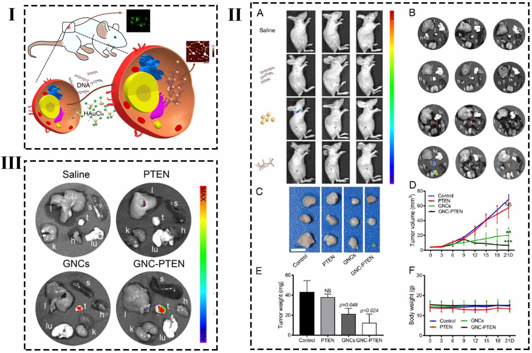
6.2. Pathological-responsive hybrid nanomaterials for phototheranostics
With the continuous development of nanotechnology, more and more nanomaterials have been widely used in the diagnosis and treatment of diseases. Exciting research results have been obtained in vitro and in vivo experiments, providing new research ideas for the treatment of major diseases (such as tumors). However, due to the small size and unique physical and chemical properties of nanomaterials, their safety and long-term performance have aroused researchers' concern. In order to realize the full clinical potential of hybrid nanomaterials, it is important to establish the interaction relationship between nanoparticles and their biological effects. Based on fully understanding the toxicity mechanism, the structure of nanoparticles can be designed more reasonably, to overcome some major obstacles in the process of their expression in vivo. The physiological response of nanoparticles can be evaluated in vitro and in vivo. In vitro evaluation of the present study mainly involves the molecular and cellular mechanisms related to nanotoxicity, and in vivo evaluation mainly involves cellular binding and durability, complement activation, oxidative stress, inflammation, DNA damage and other toxicity of nanoparticles in vivo.
In situ biosynthesis used ionic solution as the precursor, with the various enzymes in specific tissues and cells for redox reaction, to produce nanoparticles with unique properties, which can be used in the diagnosis and treatment of cancer, inflammation neurodegenerative diseases. Because these nanoparticles are spontaneously produced in biological tissues, they are likely to be highly biocompatible and less toxic, and enhanced surface area and the size and shape of the particles can also be controlled in this way. Wang's team successfully synthesized AuNCs [196], AgNCs [197] and PtNCs [212] in situ in tumor tissues by injecting metal ion solution into the tail vein, and observed the behavior status and histopathological sections of the mice after injection, and confirmed that no abnormal behavior or histopathological lesions were found in the mice. The results showed that the functional nanoparticles prepared by in situ biosynthesis had good biocompatibility and no pathological damage to normal tissues and organs.
Based on histological study, Ye et al [199] collected blood samples of mice 48 h after injection of the precursor for biochemical analysis. Compared with control group, liver and kidney function parameter (for example, blood urea nitrogen, creatine kinase, lactate dehydrogenase, serum total bile acid and creatinine and glutamate pyruvate transaminase, cholinesterase and aspartate aminotransferase) showed no obvious difference. This result is consistent with the histopathologic analysis and the mice showed no obvious behavioral or neurological abnormalities. These data suggest that this approach can safely target tumors in vivo without causing tissue cytotoxicity or abnormal physiological life function. This method can effectively avoid the cytotoxic and side effects of traditional chemical synthesis nanoparticles and has a better clinical application prospect.
The results of pathological study showed that the in situ biosynthesis of nanoparticles could only accumulate in the tumor or inflammation and would not affect the physiological functions of other normal tissues and organs, suggested that the synthesis of nanoparticles was likely to take advantage of the special physiological environment in the tumor or inflammation and participated in the physiological metabolic process. In order to further reveal the molecular mechanism of in situ biosynthesis, cDNA microarray was used to study the whole genome expression, and GO analysis and KEGG pathway analysis were performed on the differentially expressed genes. We found that in the process of in situ biosynthesis of fluorescent nanocomplexes, there are several important biological pathways involved, including the focal plaque pathway and the PI3K (phosphatidylinositol 3ʹ-kinase)-Akt signaling pathway, which can affect glycolytic metabolism and NAD(P)H-oxidase metabolism [215]. Based on genomics studies, the molecular mechanism of the differentially expressed proteins in the process of in situ synthesis in cells was further studied by using the iso-heavy isotopic multi-label relative quantitative proteomics (iTRAQ) [199]. It was found that during the co-culture of the precursor (metal ion solution) and tumor cells, ions interact with the high content of GSH in tumor cells and form fluorescent nanoparticles or nanocomplexes under the special reduction action of NADPH. The reaction process involves several special biological pathways, including the spot adhesion pathway and the phosphatidylinositol kinase signaling pathway (PI3K-Akt signaling pathway) etc, which are mutually corroborated with the genomics results. This study elucidated the mechanism of FL in tumor cells at the molecular level, providing a theoretical basis for further revealing the molecular mechanism of in situ biosynthesis of bioluminescent probes in tumors.
7. Clinical challenges of hybrid nanomedicine in vivo
The unique optical, magnetic, ultrasonic, and other properties of hybrid nanoparticles can not only be used as molecular imaging probes, but also can play an important role in tissue engineering and regenerative medicine by the optimized design of hybrid particles. For example, it can be used as a tissue scaffold to support for the body and deliver critical substances such as growth factors [216, 217]. Electrical conductivity is used to provide important electrical stimulation to heart tissue and so on [218].
Although many different types of nanomedicine have been approved clinically, many problems have been exposed with clinical trials. The targeting and metabolic pathways of nanomedicine are the two most controversial issues and have become the focus of clinical trials. Therefore, it is necessary to have a clear understanding of the challenges and difficulties in the applying of hybrid nanomaterials to solve these problems better. Here, we will focus on the effects of hybrid nanomaterials design-related factors on therapeutic outcomes.
Recent studies have shown that only about 0.7% of nanoparticles reach the tumor site when injected intravenously [219]. This inefficient delivery is not only likely to be inefficient for treatment, but also to leave large amounts of nanoparticles in the body, which may interact with tissues and organs and cause toxicity problems. Moreover, even when the nanoparticles reach their target locations, they must be able to solve problems such as tissue penetration and uniform distribution effectively. In order to better control the biological performance of nanoparticles in vivo, the fundamental parameters should be rationally designed first, such as size, surface functionalization, and so on. These physicochemical properties may directly affect its interaction with cell tissue in vivo. In addition, the cost and batch-scale synthesis factors should be considered.
7.1. Particle size of hybrid nanoparticles
Particle size is the crucial property of hybrid nanoparticles in vivo, which directly affects the drug load rate, pharmacokinetics and distribution in vivo, etc. However, different parts of the body and different diseases have different requirements on size, so it is not easy to determine a range of particle size, which needs to be changed according to specific applications.
According to the glomerular filtration cut-off value of about 5.5 nm [220], the size of nanoparticles for intravenous injection should be greater than 10 nm. Generally, phagocytosis tends to absorb particles larger than 500 nm, while clathrin-mediated and caveolae mediated endocytosis with a size of 50–500 nm [221, 222]. Sometimes, in order to make better use of the EPR effect, the size of nanoparticles is usually controlled within the range of 100–200 nm. Clinical studies have shown that without special preconditioning, the upper limit of the BBB is 10 nm. Therefore, there is a need to balance the targeted aggregation of nanoparticles with the potential for removal. Moreover, due to the high heterogeneity of tumor tissues' blood vessels, the size of hybrid nanoparticles is not always correlated with nanoparticles' accumulation at the target location. Therefore, specific design should be carried out according to the characteristics of the target location, and specific analysis should be conducted through the experimental results.
7.2. Stability of hybrid nanoparticles
The surface charge and hydrophobicity of hybrid nanoparticles determine their stability in physiological environment. In general, the amount of surface charge on nanoparticles is proportional to the stability. If the surface of nanoparticles is modified with amphoteric molecules, their stability is affected by pH. Therefore, modification of biomolecules or polymers on hybrid nanoparticles surfaces is conducive to enhanced stability [218, 223, 224]. However, it should also be noted that if the molecular weight is too high, or if the assembly is not uniform, it will cause nanoparticles to gather. The results of in vitro simulations are often quite different from those of in vivo experiments, especially for injected nanoparticles intravenously into the system. This is due to complex blood components that interact with nanoparticles.
7.3. Protein corona formed on the surface of hybrid nanoparticles
When the hybrid nanoparticles enter the body, they can easily absorb biological molecules in biological fluids (such as blood and peritoneal fluid) and form the protein corona layer [225–227]. This protein-coated structure will lead to a series of changes such as reduced surface energy and increased steric hindrance of hybrid nanoparticles, which not only dramatically conceals the intrinsic properties of the hybrid nanoparticles itself, but may also endue new physicochemical properties [228, 229]. For example, protein corona formed in the early stage affects the absorption, targeting and distribution of particles and tends to lead to hemolysis or endothelial cell death [230–232]. Therefore, for actively targeted nanomedicines, this may also be an important reason for their poor performance in clinical trials [233].
In order to reduce the negative effects of protein corona, researchers tried to change the composition, shape, surface charge and other factors of hybrid nanoparticles to avoid the formation of protein corona [234–236]. In addition, it should be noted that the disease variability and heterogeneity, as well as individual differences of patients, lead to different effects of protein corona [237]. We also noticed that the role of protein corona is not necessarily negative [238, 239]. Therefore, it is more necessary for researchers to make in-depth and comprehensive explorations in related fields, including biological interface behavior.
7.4. Targeted of hybrid nanoparticles
Precise control of the release of nano-drugs in target tissues and organs is an important challenge to translate into clinical applications. In some ways, utilization and targeting are similar concepts. By modifying ligands on the surface of particles, the biocompatibility and distribution in vivo can be effectively regulated, so that drug-loaded nanoparticles can accumulate in large quantities on the target tissue and thus avoid toxicity to other organs.
For oral nanodrugs, the effects on gastric mucosa, intestinal cells, and liver should be considered according to their absorption and metabolic pathways. At present, various cellular mechanisms, such as receptor-mediated endocytosis, phagocytosis and so on, have been used to improve the utilization rate of oral preparations [240, 241]. To design nanoparticles for the treatment of brain diseases, it is necessary to fully investigate the barrier effect of BBB on nanoparticles. The researchers used intranasal administration or preconditioning (e.g. opening the BBB in advance, using transport mechanisms) to achieve BBB penetration [242]. Different modes of administration, such as intraperitoneal injection [243, 244] and percutaneous injection [245, 246], need to overcome different barriers or internal environments. Therefore, researchers are required to carry out targeted design according to the actual needs.
7.5. Other aspects
In addition, small amounts of nanoparticles produced in laboratories are often controllable and of better quality. However, the number of nanoparticles required in quasi-clinical and clinical trials is much higher than the scale of synthesis in the laboratory, so the production of nanoparticles needs to be expanded on an industrial scale. In the actual operation process, it was found that the expanded production could easily lead to many problems, such as uneven particle size, reduced stability, original retention, and different quality between batches. These factors directly affect the properties of nanoparticles and may cause significant changes in the biological behavior of nanoparticles. Therefore, researchers need to take these factors into account when preparing nanoparticles on a large scale. In addition to controlling the quality of nanoparticles, cost control is also of particular importance in order to further promote drug-carrying nanomaterials in clinical practice. For example, the US FDA proposed a quality design method, which can well control the cost of materials and effectively control the factors that may affect the safety and quality of drugs [247].
As discussed in the previous sections, so far, a great deal of effort and funds have been invested in the development of hybrid nanomaterials in preclinical and clinical trials, and great progress has been made. However, to achieve clinical use and industrial mass production, there are still many obstacles to overcome. In addition to adjusting and improving various parameters of hybrid nanomaterials themselves, it is necessary to fully combine these properties with the internal environment of the human body and individual differences of patients, to find a delicate balance to meet the needs of clinical transformation.
First, an important criterion for the clinical application of biomedicine is safety, efficiency and controllability. Therefore, it is required that the design and synthesis of hybrid nanomaterials should be as simple as possible. However, the current design of hybrid nanomaterials is usually too complicated. Although the preparation of hybrid nanomaterials by in situ biosynthesis is relatively simple, the purification of materials is more difficult and the yield is not high. Secondly, to explore how hybrid nanomaterials can function in the human body, it is necessary to fully understand the material's biodistribution, potential degradation, removal, and biocompatibility in the body. It is even necessary to explore the interaction between hybrid nanomaterials and proteins, cells, and tissues at the molecular level, reduce their distribution in normal tissues, and effectively reduce toxic and side effects. These studies are of great significance for real clinical applications, but they also encounter great difficulties at present. For example, in FL research, due to the complex composition of blood, the material will cause damage to its surface after entering the blood, and blood absorption will weaken its signal strength. Due to the poor penetration of fluorescence, the detection of thick bone tissue and deep tumors is still challenging. In the treatment research, repeated demonstrations and long-term follow-up studies are required for the drug loading dose, drug release mode, and drug residue status of hybrid nanomaterials.
The advantage of nano-hybrid materials is that they can aggregate and synergize the performance and functions of individual components to achieve the greatest optimization effect. With the continuous innovation of synthetic technology and the in-depth understanding of biological systems, more simple and new design concepts are developed, which may be more practical. Nano-hybrid technology for disease imaging and imaging-guided treatment research will receive more attention, because it can realize real-time and long-term monitoring of the disease treatment process while realizing early and accurate diagnosis. Advances in synthesis technology will inevitably reduce the toxicity to biological systems, making hybrid nanomaterials feasible in clinical applications. We believe that with the continuous in-depth research on hybrid nanomaterials, it is expected that the materials can be used to provide patients with new treatment plans and more personalized treatments.
Acknowledgments
This work was financially supported by the National Natural Science Foundation of China (82061148012, 82027806, 91753106), Primary Research and Development Plan of Jiangsu Province (BE2019716), and the National High Technology Research and Development Program of China (2017YFA0205300).
Data availability statement
No new data were created or analyzed in this study.