

Abstract
Pharmacokinetics of nanomedicines can be improved by a temporal blockade of mononuclear phagocyte system (MPS) through the interaction with other biocompatible nanoparticles. Liposomes are excellent candidates as blocking agents, but the efficiency of the MPS blockade can greatly depend on the liposome properties. Here, we investigated the dependence of the efficiency of the induced MPS blockade in vitro and in vivo on the size of blocking liposomes in the 100–500 nm range. Saturation of RAW 264.7 macrophage uptake was observed for phosphatidylcholine/cholesterol liposomes larger than 200 nm in vitro. In mice, liposomes of all sizes exhibited a blocking effect on liver macrophages, prolonging the circulation of subsequently administrated magnetic nanoparticles in the bloodstream, reducing their liver uptake, and increasing accumulation in the spleen and lungs. Importantly, these effects became more pronounced with the increase of liposome size. Optimization of the size of the blocking liposomes holds the potential to enhance drug delivery and improve cancer therapy.

Original content from this work may be used under the terms of the Creative Commons Attribution 4.0 license. Any further distribution of this work must maintain attribution to the author(s) and the title of the work, journal citation and DOI.
1. Introduction
Nanoparticles are commonly employed for medical treatment as drug delivery vehicles and contrast agents. However, the systemic administration of nanomedicines is often associated with unfavorable uptake by liver, limiting their accumulation in other tissues. The high particle sequestration by the liver is primarily attributed to the activity of Kupffer cells, which constitute the major cell population within the mononuclear phagocyte system (MPS) [1]. Macrophages in the spleen, lungs, bone marrow and other tissues also contribute to the phagocytic activity of the MPS, but their overall capacity for nanoparticle uptake is limited [2].
To prevent non-specific liver uptake, several strategies of particle engineering are commonly employed, including shielding from immune cell uptake through stealth coating [3] and functionalization with biochemical moieties like self-peptides [4]. However, these approaches still have several drawbacks, including immune reactions to the polymer chains, hampered nanoparticle interaction with target cells and insufficient increase of the delivery to tumour [5].
Other approaches for improved drug delivery aim to affect the MPS itself [6]. Clodronate and propamidine delivered to the liver and spleen deplete macrophage populations by inducing mitochondrial disfunction and consequent apoptotic cell death [7]. Another group of small molecules inhibits the endocytic pathways responsible for the uptake of nanoparticles. For instance, the treatment of Kupffer cells with chloroquine is known to suppress expression of phosphatidylinositol binding clathrin assembly protein, which is critical for clathrin-mediated internalization [8, 9]. GdCl3 and zoledronic acid are other inhibitors used to inactivate clathrin-independent nanoparticle uptake [9, 10]. Recently, genetic downregulation of endocytic receptors of macrophages was proposed for improving exosome delivery [11].
An alternative strategy is MPS-blockade, which suppresses the endocytic activity of cells after the massive uptake of low-toxic nanomaterials [12, 13]. Subsequently administrated nanomedicines can bypass non-specific uptake in macrophage-rich tissues, leading to prolonged blood circulation kinetics. The MPS blockade can be achieved after intravenous administration of high doses of various low-toxic nanomaterials, such as polymeric [14], lipid [15], inorganic [16] or protein nanoparticles [17]. For instance, we demonstrated that the magnetic field assisted targeting, in combination with MPS blockade by ferrihydrite nanoparticles, increased the delivery of magnetic nanoparticles (MNPs) to the tumor from ∼0.2% to more than 6% of the injected dose (ID) [2]. While solid inorganic nanomaterials are capable to induce strong MPS blockade [16], their slow metabolism limits safe application of the method.
Unlike many biodegradable inorganic blocking nanoparticles that are currently under development [14, 18], liposomes have a long history of clinical use as drug carriers and are known for their high biocompatibility [19]. MPS blockade with liposomes transiently prolongs the circulation of therapeutic and diagnostic nanoparticles in the bloodstream, as well as improves the tumor delivery efficacy [12, 20]. Liposomes did not induce liver and spleen toxicity or inhibit MPS-mediated host defense against bacteria at a high dose of 376 mg kg−1 [12]. The approach efficiency can be reinforced by increasing the dose of injected liposomes, or by using positively charged blockers [21]. The direct comparation of ultrasmall (20–50 nm) and large (650–2000 nm) liposomes showed that the larger ones induce blockade faster, which is explained by their rapid clearance (Cl) from circulation [22].
In this study, we investigated the effect of liposome size within the range of 100–500 nm on MPS blockade efficiency, both in vitro and in vivo. We utilized liposomes composed of phosphatidylcholine/cholesterol (PC/Chol), which demonstrated low toxicity to macrophages even at high doses. Large liposomes reduced uptake of iron oxide nanoparticles in RAW264.7 macrophages, while liposomes smaller than 200 nm had no discernible effect. In vivo analysis of iron oxide nanoparticle circulation after the MPS blockade showed higher macrophage saturation with large liposomes. The blockade also induced a significant redistribution of particles from liver to spleen and lungs. These findings provide valuable insights for the development of therapies involving the induction of the liposomal MPS blockade.
2. Results and discussion
2.1. Preparation of liposomes
We prepared liposomes composed of L-α-PC and Chol, common components of clinically approved lipid-based therapeutics [23]. While non-PEGylated PC/Chol liposomes are used for drug delivery and tumor treatment [24], liver and spleen remain main organs of their accumulation in the body [25, 26]. Pre-dosing with liposomes of PC/Chol composition has been reported to deplete the MPS activity by occupation of the hepatic and splenic phagocytes [27]. Moreover, the high biocompatibility and cost-effectiveness of these lipids make them attractive for preparing MPS-blocking liposomes, especially considering the typically high doses required for an effective blocking effect in vivo (∼100 mg kg−1 of body weight) [12, 14, 28].
Nanosized PC/Chol (1/1 molar ratio) liposomes were synthesized via a thin-film hydration method, followed by membrane extrusion-based size fractionation. We produced liposomes larger 100 nm to avoid renal excretion, while liver and spleen uptake dominates for liposomes of this size [25, 29].
Three size fractions of liposomes were employed to induce MPS-blockade, with mean hydrodynamic diameters of 160 nm (small, LPS), 210 nm (medium, LPM) and 370 nm (large, LPL), each having low polydispersity (PDI < 0.25). The size distribution and corresponding dispersity parameters are presented in figure 1(a) and supplementary table 1. All formulations demonstrated stability of the mean hydrodynamic size in PBS over a period of 8 h (figure 1(b)). The size of all liposomal fractions was higher than endothelial fenestrae in liver, preventing these nanoparticles from entering the space of Disse and increasing the probability of interaction with Kupffer cells [30]. Nanosized PC/Chol liposomes possessed a strong negative ζ-potential in 10 mM NaCl (LPS: −43 ± 9 mV, LPM: −30 ± 10 mV, LPL: −34 ± 7 mV), indicating similar surface properties and high colloidal stability of nanoparticles (figure 1(c)). The negative surface charge of liposomes composed of zwitterionic PC lipid and Chol is consistent with other studies [31] and can be explained by high affinity of the PC chains to anions in salt solution.

Figure 1. Characterization of blocking liposomes. (a) Hydrodynamic size distribution and (b) evolution of mean hydrodynamic diameter of small (LPS), medium-sized (LPM) and large (LPL) liposomes in PBS over time. (c) ζ-potential analysis of liposomes. (d) MTT cytotoxicity test of liposomes towards RAW264.7 macrophages after 24 h of co-incubation. n = 3. Data are presented as mode ± SD of distribution (b), (c) or mean ± SD (d).
Download figure:
Standard image High-resolution image2.2. Biocompatibility of liposomes to macrophages
Macrophages represent a target cell population in MPS-blockade approaches, as they are responsible for elimination of major fraction of exogeneous nanomaterials in vivo [32, 33]. Therefore, it is important to access cytotoxicity of liposomal blockers towards these cells. For this aim, we utilized the commonly used murine macrophage RAW264.7 line and measured their viability by MTT test. The experiment was conducted under high concentrations of liposomes (up to 500 μg ml−1), which are expected to saturate macrophage uptake function in vitro [21, 28].
For all three types of prepared liposomes, a gradual decrease in viability was observed with an increase in particle concentration (figure 1(d)). No significant difference in cytotoxicity was found between liposomes of the studied size ranges. At a concentration of 50 μg ml−1, the viability was approximately 85%, and after a 10-fold dose increase it plateaued at the 70%–73% level. The mild toxicity of liposomes may be explained by the presence of remnants of organic solvents in the particles structure [34]. Nevertheless, nanosized liposomes demonstrate a low level of cytotoxicity to macrophages, compared to many other nanoparticles, used for the MPS-blockade. For example, silica (IC50 < 600 μg ml−1 [35]) and polystyrene beads (IC50 < 500 μg ml−1 [36]) are found to be toxic at high doses. For the designed liposomes, the IC50 was higher than 500 μg ml−1 and could not be determined from the graphs. Thus, the PC/Chol liposomes of the 100–500 nm size range have low toxicity to macrophages even at high doses.
2.3. Macrophage blockade in vitro
Blocking nanoparticles are designed to inhibit endocytosis of macrophages via temporal occupation and saturation of their surface receptors [12, 28]. Therefore, we compared uptake of MNPs in macrophages after their blockade with LPS, LPM and LPL formulations. MNPs are commonly applied in nanomedicine for therapeutic and diagnostic purposes, but their rapid elimination from circulation by professional phagocytes remains a challenging problem [37]. In this study, we used multicore magnetic iron oxide nanoparticles stabilized by glucuronic acid (FluidMag-ARA) as model of therapeutic nanoparticles. They have hydrodynamic diameter of (160 ± 40) nm and ζ-potential of (−34 ± 7) mV (figures S1(a) and (b)). Nanoparticles of similar size are widely employed for drug delivery due to their sufficient payload loading, avoidance of renal filtration, and potential for their retention in tumors via EPR effect [38]. Nevertheless, rapid macrophage uptake hampers their performance. Superparamagnetic properties of FluidMag-ARA nanoparticles allow to quantify them in samples and tissues by the magnetic particle quantification (MPQ) technique [39].
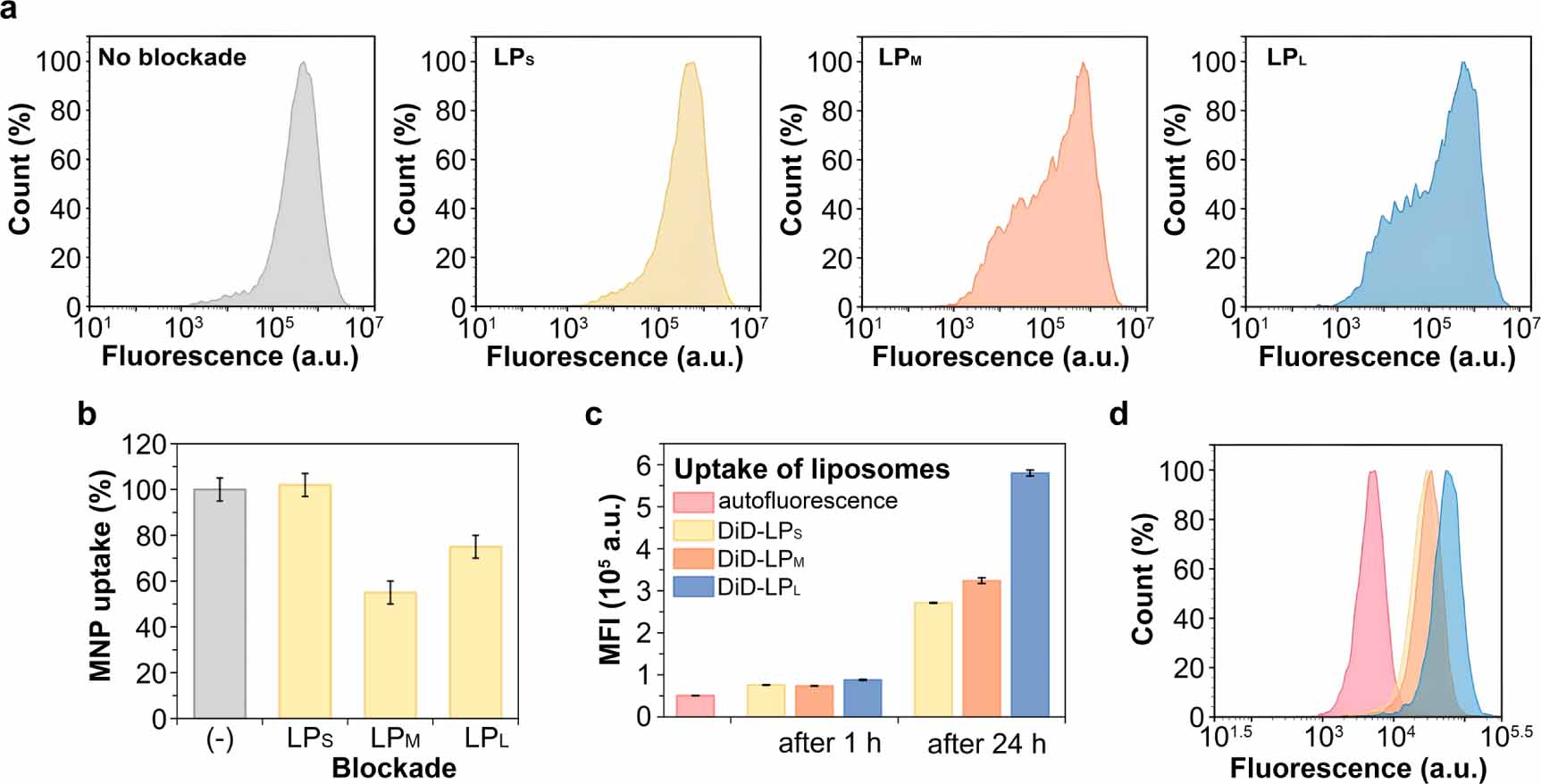
The liposomal blockade in vitro was performed using RAW264.7 macrophages. Liposomes were added to the cells at a concentration of 500 μg ml−1 and incubated for 24 h to saturate endocytic pathways. The cells were then washed with PBS to remove the unbound liposomes, and MNPs labeled with a lipophilic fluorescent dye (FluidMAG-ARA/R) were added to the cells at 25 μg ml−1 dose and co-incubated for 24 h. The fluorescent MNPs had similar size and ζ-potential distributions as non-labeled ones (figures S2(a) and (b)). Cellular uptake of MNPs was accessed by flow cytometry (figure 2(a)) and compared by the distribution of cell fluorescence intensity (figure 2(b)). The blockade of macrophages was manifested when cells were incubated with LPM and LPL formulations. Flow cytometry histograms shifted to the smaller fluorescence values 24 h after the blockade (figure 2(a)), and the calculated uptake of MNPs decreased by 45% and 25% after the saturation with LPM and LPL, respectively (figure 2(b)). However, after the treatment of macrophages with small-sized LPS liposomes, no significant change in the uptake of MNPs was observed.

Figure 2. Macrophage blockade in vitro. (a), (b) Flow cytometry analysis of fluorescent MNPs uptake in RAW264.7 cells 24 h after blockade with different liposomal formulations. The histograms (a) were acquired in YL2 channel to measure MNPs uptake by median fluorescence intensity (MFI) values (b). (c), (d) Flow cytometry analysis of DiD-labelled blocking liposomes uptake by RAW264.7 cells after 1 h (c) and 24 h (c), (d) incubation; the histograms are acquired in YL3 channel. In (b), (с) data are presented as mean ± SD, n = 3.
Download figure:
Standard image High-resolution imageObserved difference may be explained by the kinetics of liposomal uptake by cells. To test this assumption, we synthesized liposomal formulations of similar size distributions labeled with a fluorescent 1,1'-dioctadecyl-3,3,3',3'-tetramethylindodicarbocyanine (DiD) dye (see characterization at table S1 and figure S3). Labeled liposomes were added to RAW264.7 macrophages at an equal lipid dose (500 μg ml−1), incubated for 1 h or 24 h, and analyzed by flow cytometry for DiD fluorescence. Macrophages showed similar uptake of all types of liposomes at 1 h time-point (figure 2(c)). However, after incubation for 24 h, DiD-LPM and DiD-LPL demonstrated higher cellular uptake with a 20% and 110% increase in median fluorescence intensity compared to DiD-LPS, respectively (figures 2(c) and (d)). Low accumulation of LPS in cells was additionally confirmed by fluorescence microscopy (figure S4). Significant difference in the blockade efficiency by LPS and LPM formulations cannot be explained by the liposomal uptake alone, and other factors, such as the speed of receptor recycling and rate of endocytosis, are likely involved.
2.4. Pharmacokinetics of blocking liposomes
Pharmacokinetics of medium-sized liposomes was determined to understand time delay between blocking and magnetic particle administration. For this aim, fluorescent DiD-labeled liposomes of intermediate size (DiD-LPM) were administered in BALB/c mice at a dose of 3 mg via retro-orbital injection. Their blood availability was evaluated by measuring the fluorescence of plasma samples (figure 3(a)). The pharmacokinetics exhibited a biexponential profile with half-lives of (13.2 ± 2.5) min and (3.7 ± 2.3) h for the rapid and slow phases, respectively. Within the first hour of circulation, the signal of fluorescence in blood dropped 2.2-fold. After 1 h, spleen and liver demonstrated the strongest fluorescence among organs (figure 3(b)), confirming effective capture of liposomes by resident macrophages. For the next 6 h, the fluorescence of spleen decreased by 55%, while the liver retained the same signal. From this study, we conclude that 1 h is a sufficient interval for MPS blockage in vivo with PC/Chol liposomes, considering that the liver has substantial uptake of liposomes, and part of the blocking materials remains in the bloodstream to compete with exogenous nanoparticles for recognition [40].

Figure 3. Pharmacokinetics of blocking liposomes. (a) Blood circulation kinetics of 200 nm liposomes labeled with fluorescent dye (DiD-LPM) and (b) their biodistribution. Data are shown as mean ± SD. n = 3.
Download figure:
Standard image High-resolution image2.5. Macrophage blockade in vivo
To evaluate the circulation kinetics of MNPs in the bloodstream and their biodistribution in mice, we utilized the MPQ technique, described in detail by Zelepukin et al [39]. The MPQ method quantitatively and non-invasively measures concentration of non-linear magnetics, such as superparamagnetic nanoparticles, in the blood vessels of mouse tail. Linear paramagnetic and diamagnetic materials, such as animal tissue and iron in the blood, do not contribute to the measured signal, setting the limit of detection of nanoparticles at the nanogram level. This method enables real-time tracking of pharmacokinetics of magnetic particles with high sensitivity.
Macrophage blockade was induced by injection of 3 mg of liposomes into the orbital venous sinus followed by administration of MNPs after 1 h via the same route. MNPs blood circulation kinetics and biodistribution at 3 h were analyzed (figure 4(a)). In the absence of blockade, the elimination kinetics of MNPs exponentially decreased and 90% of the ID was cleared within 7 min (figure 4(b)). MPS blockade with liposomes significantly prolonged the circulation of nanoparticles. The half-life increased from (1.2 ± 0.1) min to (6.2 ± 2.1) min, (7.0 ± 2.7) min and (7.5 ± 2.8) min using LPS, LPM and LPL blockers, respectively. Other common parameters of pharmacokinetics analysis, such as area under curve and Cl demonstrated the beneficial effect of blockade on the prolongation of MNP circulation (table S2). Furthermore, the highest values were observed for LPL blockers, as consistent with the in vitro study. Nevertheless, LPS nanoparticles, which were completely inefficient in vitro, showed in vivo blockade effect similar in magnitude to LPM nanoparticles.

Figure 4. (a) Scheme of the macrophage blockade experiment with liposomes for modulating pharmacokinetics of MNPs. Mice were injected into retroorbital venous sinus with a blocking dose of LP 1 h prior to administration of MNPs. The circulation kinetics of MNPs was recorded in tail veins and after 3 h, biodistribution of MNPs was analyzed. (b) Effect of liposomal blockade on blood circulation kinetics of MNPs. Insert—kinetics plotted in logarithmic scale. (c)–(e) Biodistribution of MNPs by organs dependent on blockade with different liposome formulations. (h) Ratio of MNPs accumulated in liver to spleen. Data are presented as mean ± SD. n = 3 animals per condition. (*)—p < 0.05, (**) p < 0.01, two-tailed Welch's t-test.
Download figure:
Standard image High-resolution image
This difference can be explained by the involvement of other saturation mechanism in complex in vivo environment. For instance, depletion of opsonins in serum by liposomes can additionally decrease the subsequent uptake of MNPs [12, 41]. In addition, small liposomes generally demonstrate longer blood circulation time and can better compete for binding to macrophage surface during MNPs circulation [40].
The biodistribution of MNPs after liposomal blockade is presented in figures 4(c)–(h) and S5, raw data are summarized in table S3. Without the blockade induction, 99% of the ID accumulated in liver and spleen, two major organs of MPS. Pre-treatment with liposomes significantly decreased total accumulation in these organs by 8.5%, 10%, and 6.6% ID for LPL, LPM, and LPS liposomes, respectively. Low effect of LPS liposomes on depleting activity of the liver and spleen phagocytes corroborates with the observation on the lower uptake by macrophages in vitro. The delivery of MNPs to the lungs increased in order of magnitude after the blockade with all three blocker types, but the effect was most pronounced for the largest LPL liposomes. In addition, the blockade with LPM and LPL liposomes induced a 3-fold increase in delivery to the kidneys and 4.3- and 3.7-fold higher uptake in the heart, respectively. At the same time, LPS liposomes did not change significantly the uptake of MNPs in these organs compared to the non-blocking condition (table S3).
Importantly, even after the liposomal blockade, liver remains the main organ eliminating nanoparticles from the bloodstream. Kupffer cells in the liver constitute 80%–90% of the total macrophage population in the body [32, 42], and they reside in sinusoids with slow blood flow, which increases the probability of nanoparticle capture [1]. Tavares et al reported that even after elimination of 75% of liver macrophages by clodronate liposomes, the liver continues to be the second organ of nanoparticle uptake after the spleen [43]. MPS blockade with liposomes is a mild method and only partially and temporally reduces the activity of the liver macrophages, so magnetic particles still accumulate in this organ in high quantities.
Nevertheless, since liver serves as the first barrier encountered by nanoparticles after the injection, MPS blockade induces a 'spillover effect' of nanoparticle redistribution to other macrophage-reach tissues like spleen and bone marrow [44]. The improvement of the splenic uptake of nanomedicines is currently desirable for gene delivery and immunization applications, as well as for direct treatment of spleen pathologies [45, 46]. Therefore, we further examined difference in distribution between liver and spleen dependent on blocking liposome size (figures 4(c), (d) and (h)). The average liver accumulation of MNPs was observed to decline gradually with the increase in liposome size, from 78.6% ID (LPS) to 71.6% ID (LPL). On the other hand, the average quantity of MNPs reaching spleen was highest for LPL formulation (18.5% ID), with no significant difference observed between blockade by LPM and LPs formulations (∼14% ID). The ratio of liver to spleen accumulation of magnetic particles decreased from 7.9 in animals without blockade to 3.9 in mice with blockade by LPL liposomes. Decreased liver-to-spleen delivery ratios were established in LPS and LPM groups, with corresponding values of 5.8 and 5.3. Our observations suggest that the increase of mean liposome size towards 370 nm provides higher splenic uptake of post-injected therapeutic nanoparticles by hindering their capture by liver. It corroborates with recent meta-analysis of MPS blockade studies, where the spillover effect was observed to increase for nanomaterials larger 200 nm [6]. However, further size increase of blocking liposomes does not guarantee an elevation of liver-evasion capacity, since the hepatic uptake dependence for liposomes typically demonstrates saturation at 250–400 nm mean liposome sizes [36].
3. Conclusion
Liposomal MPS blockade has been widely employed to enhance tumor delivery and to prolong the blood circulation of therapeutic and diagnostic nanoparticles. However, many approaches saturate macrophages using lipid emulsions with high polydispersity. In this study, we evaluated the influence of the size of liposomes on the effectivity of induced macrophage blockade both in vitro and in vivo. All three fractions of liposomes with mean hydrodynamic diameters of 160, 210, and 370 nm exhibited low toxicity towards the RAW264.7 macrophage cell line. However, only liposomes larger than 200 nm induced blockade in vitro, leading to a decrease in uptake of subsequently introduced magnetic particles. Large liposome fraction also induced a stronger blockade in vivo, as confirmed by the greater prolongation of magnetic particles blood circulation and their higher redistribution from the liver to other macrophage-rich and normal tissues. This effect can be partially explained by the higher uptake of large-sized liposomes by macrophages. Nevertheless, even liposomes smaller than 200 nm induced blockade in vivo, revealing an inconsistency between in vivo and in vitro data and underscoring the importance of optimizing macrophage blockade in animal studies. The obtained results can contribute to the rational design of nanoparticles for the MPS blockade, wherein larger nanoparticles with low polydispersity will play a leading role.
4. Materials and methods
4.1. Materials
L-α-PC, Chol and methanol were obtained from Sigma-Aldrich, USA. Chloroform and dimethyl sulfoxide (DMSO) were obtained from Chimmed, Russia. 3-(4,5-dimethylthiazol-2-yl)-2,5-diphenyl-2H-tetrazolium bromide (MTT) and 1,1-dioctadecyl-3,3,3,3-tetramethylindodicarbocyanine (DiD) were obtained from Thermo Fisher Scientific, USA. FluidMAG-ARA100 and FluidMAG-ARA100/R nanoparticles were purchased from Chemicell GmbH, Germany. L-glutamine, penicillin, streptomycin, and amphotericin B were purchased from Gibco, USA.
4.2. Synthesis of liposomes
56 mg of L-α-PC and 28 mg of Chol were dissolved in 5 ml of 6:1 chloroform/methanol solution and then dried under reduced pressure using a rotary evaporator at 40 °C and 90 rpm rotation speed. The obtained lipid film was hydrated with PBS (0.1 M, pH 7.4) in an ultrasonic bath. Microscale liposomes were collected by centrifugation at 100 g for 5 min. Then, liposomes were extruded through 400 nm, 200 nm and 100 nm polycarbonate membranes to obtain liposomes of the desired sizes.
To prepare fluorescent liposomes, DiD dye was added to chloroform/methanol mixture in a weight ratio of 0.01% of a total lipid mass. After extrusion, liposomes were washed from the unbound dye by centrifugation in spin columns with 7 kDa weight cut-off.
4.3. Characterization of nanoparticles
The size distribution and ζ-potential of liposomes and MNPs were characterised using Zetasizer Nano ZS device (Malvern, UK). The hydrodynamic size (diameter) of liposomes was determined from intensity size distribution. ζ-potential of liposomes and MNPs was measured in 10 mM NaCl. The Smoluchowski approximation was used for analysis.
4.4. Cells
RAW264.7 (ATCC TIB-71™) macrophage line were cultured in DMEM medium supplemented with 10% FBS, 2 mM L-glutamine and Gibco antibiotic–antimycotic solution. Cells were seeded in 96-well plates at density of 104 cells per well and incubated at 37 °C under humidified atmosphere containing 5% CO2 for 24 h before the experiments.
4.5. Cell viability
Macrophage cells were co-incubated with liposomes at final concentrations of 0.5, 0.1 and 0.05 mg ml−1 in 100 μl of medium. After 24 h of incubation, the medium was removed and 100 μl of the MTT solution (0.5 g l−1 in DMEM) was added to each well. The cells were incubated for 1 h at 37 °C, MTT solution was removed and 100 μl of DMSO was added to the wells. Then the plate was shaken in the dark until complete dissolution of formazan crystals. Cell viability was measured by optical density at 570 nm wavelength, using Infinite 1000 PRO (Tecan, Austria) microplate reader, and was normalized to the viability of untreated cells.
4.6. Macrophage blockade in vitro
Liposomes at concentration of 0.5 mg ml−1 were incubated with RAW264.7 macrophages for 24 h. Then, cells were washed with PBS and treated with 25 μg ml−1 of FluidMAG-ARA100/R fluorescent nanoparticles in DMEM medium (excitation: 578 nm, emission: 613 nm). In 3 h, cells were washed 3 times with 1% BSA in PBS and analyzed by a Novocyte 3000 cytometer in the YL2 channel (excitation laser at 561 nm, emission filter at 615/20 nm). The uptake of fluorescent nanoparticles after liposomal blockade was determined by median intensity of cell fluorescence and was normalized to the uptake values in cells without blockade.
To visualize liposomal blockade of macrophages, DiD-labelled liposomes were added to the cells to a final concentration of 0.5 mg ml−1. In 1 h or 24 h, the cells were washed with 1% BSA in PBS and analyzed by flow cytometry in the YL3 channel (excitation laser at 561 nm, emission filter at 660/20 nm), and by fluorescence microscopy on a DMI 6000B microscope (Leica, Germany) equipped with a DFC295 camera.
4.7. Animals
All experiments with animals were approved by the institutional animal care and use committee of Shemyakin–Ovchinnikov Institute of Bioorganic Chemistry Russian Academy of Sciences (IBCh RAS), protocol 367/2022 from 15.12.2022. The procedures were performed according to Institutional guidelines of IBCh RAS, based on European convention on protection of vertebrate animals (ETS 123 from 18.03.1986) and Russian animal legislation. Female BALB/c mice of 18–22 g (10–14 weeks old) were obtained from Pushchino animal facility (Pushchino, Russia) and maintained at the vivarium of IBCh RAS. Animals were maintained under 12 h dark-light cycle with a constant supply of the food and water. Before nanoparticles administration, mice were anesthetized with intraperitoneal injection of Zoletil/Rometar mixture at 53 and 2 mg kg−1 doses, respectively.
4.8. Liposomes pharmacokinetics study
To study the blood circulation of liposomes, 3 mg of DiD-tagged liposomes in 300 μl of PBS (pH 7.4) were injected into retro-orbital sinus of BALB/c mice. Blood samples (30 μl) were collected with heparin within the following 7 h via puncture of the opposite retro-orbital sinus and diluted with 120 μl of PBS. Blood plasma was separated by centrifugation of samples at 1000 g for 3 min. Liposome concentration in samples was measured by fluorescence intensity at 666 nm with excitation at 647 nm on an Infinite M1000 microplate reader.
To study the biodistribution of liposomes, mice were sacrificed by cervical dislocation and major organs were analyzed by averaged radiance efficiency of epifluorescence using IVIS Spectrum CT device (PerkinElmer, USA) with excitation at 647 nm.
4.9. Macrophage blockade in vivo
3 mg of liposomes were injected into retro-orbital venous sinus of mice to induce macrophage blockade. Then, in 1 h, 300 μg of FluidMAG-ARA MNPs were injected into opposite retroorbital sinus. Magnetic particle pharmacokinetics was measured quantitatively by MPQ technique [39]. Briefly, mice tail was placed into a coil of the MPQ detector and gently fixed with a tape lent. Nanoparticle concentration in tail veins and arteries was measured with a frequency of 15.2 Hz.
Biodistribution of MNPs was measured ex vivo with MPQ technique 3 h after their administration. Mice were euthanized by cervical dislocation and major organs were extracted and placed into the coil of the MPQ detector. The measured magnetic signals were normalized to the total detected dose of nanoparticles in all extracted organs and presented in % of ID.
Acknowledgment
The work was supported by the Ministry of Science and Higher Education of the Russian Federation, Agreement No. 075-15-2024-536.
Data availability statement
All data that support the findings of this study are included within the article (and any supplementary files).
Conflict of interest
P.I.N. is a named inventor on MPQ-related patents (RU2166751, RU2177611 and others).
Authors contribution
Iaroslav B. Belyaev: Conceptualization, Investigation, Writing—original draft. Aziz B. Mirkasymov: Investigation, Writing—review & editing. Vladislav I. Rodionov: Investigation. Julia S. Babkova: Investigation, Writing—review & editing. Petr I. Nikitin: Methodology. Sergey M. Deyev: Funding, Writing—review & editing, Supervision. Ivan V. Zelepukin: Conceptualization, Supervision, Writing—original draft.