
Abstract
The Compton camera can simultaneously acquire images of multiple isotopes injected in a body; therefore, it has the potential to introduce a new subfield in the field of biomedical imaging applications. The objective of this study is to assess the ability of a prototype semiconductor-based silicon/cadmium telluride (Si/CdTe) Compton camera to simultaneously image the distributions of technetium (99mTc)-dimercaptosuccinic acid (DMSA) (141 keV emission) and 18F-fluorodeoxyglucose (FDG) (511 keV emission) injected into a human volunteer.
99mTc-DMSA and 18F-FDG were injected intravenously into a 25-year-old male volunteer. The distributions of 99mTc-DMSA and 18F-FDG were simultaneously made visible by setting a specified energy window for each radioisotope. The images of these radiopharmaceuticals acquired using the prototype Compton camera were superimposed onto computed tomography images for reference.
The reconstructed image showed that 99mTc-DMSA had accumulated in both kidneys, which is consistent with the well-known diagnostic distribution determined by clinical imaging via single-photon emission computed tomography. In the 18F-FDG image, there is broad distribution around the liver and kidneys, which was expected based on routine clinical positron emission tomography imaging.
The current study demonstrated for the first time that the Si/CdTe Compton camera was capable of simultaneously imaging the distributions of two radiopharmaceuticals, 99mTc-DMSA and 18F-FDG, in a human body. These results suggest that the Si/CdTe Compton camera has the potential to become a novel modality for nuclear medical diagnoses enabling multi-probe simultaneous tracking.
Export citation and abstract BibTeX RIS

Original content from this work may be used under the terms of the Creative Commons Attribution 3.0 licence. Any further distribution of this work must maintain attribution to the author(s) and the title of the work, journal citation and DOI.
1. Introduction
In recent times, the use of Compton cameras has been widely investigated for medical applications, such as scintigraphy, single-photon emission computed tomography (SPECT) (Nakamura et al 2016, Vernekohl et al 2016, Fontana et al 2017, Muñoz et al 2017, 2018), and range verification in hadron therapy (Ortega et al 2015, Hilaire et al 2016, Solevi et al 2016, Rohling et al 2017, Draeger et al 2018, Huang et al 2018, Parajuli et al 2019). Compton cameras reconstruct the distributions of gamma-ray-emitting radioisotopes (RI) based on Compton scattering information recorded by a detector. Figure 1(a) shows a schematic diagram of the working principle of a Compton camera. Because Compton interaction primarily occurs in the energy band ranging from 100 keV to 1–2 MeV, Compton cameras can be used in this wide energy band. Compton cameras have the potential for application in dual-energy or multi-energy RI tomography, which remains a challenging task among conventional nuclear imaging modalities, such as positron emission tomography (PET) and single photon emission computed tomography (SPECT). However, only recently has a Compton camera with sufficient angular resolution and energy resolution been developed to be suitable for the abovementioned applications.
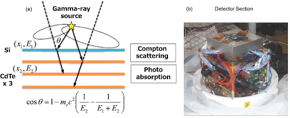
Figure 1. (a) Operation principle of Compton camera. (b) External appearance of a detector section of Compton camera in this study.
Download figure:
Standard image High-resolution imageA silicon/cadmium telluride (Si/CdTe) Compton camera was recently developed based on high-resolution CdTe semiconductor imaging devices (Takahashi et al 2004, 2012, Watanabe et al 2005, 2014, Takeda et al 2012). The Si/CdTe Compton camera can simultaneously detect technetium (99mTc), the most frequently used RI in nuclear medicine diagnosis, as well as positron emitters, such as 18F and 11C. Compton cameras are capable of using multiple RIs, which allows simultaneous imaging and shortens medical examination times by removing the need to wait for the elimination of RIs used for previous a diagnosis to avoid crosstalk. Furthermore, using the Compton camera, the reciprocal actions and metabolism of several related substances can be analyzed over time in vivo.
Multi-tracer imaging is still challenging, but it has the potential for clinical and molecular applications. For example, hypoxia is an important factor for tumor treatment. If blood flow could be observed simultaneously with hypoxia, the detection would be more precise (Lehtiö et al 2003). Multi-tracer SPECT is helpful in the differential diagnosis of parkinsonism (O'Brien et al 2014). To investigate the metabolism, a multi-tracer screening system incorporating a Compton camera has been developed (Kanayama et al 2005). Thus, the clinical application of Compton cameras is a promising advancement in nuclear diagnostic medicine. Nevertheless, several issues need to be addressed before clinically applicable Compton images can be routinely used.
The objective of the current study was to assess the efficacy of the Compton camera to simultaneously capture the distributions of 99mTc-dimercaptosuccinic acid (DMSA) (141 keV emission) and 18F-fluorodeoxyglucose (FDG) (511 keV emission) injected into a human volunteer. This study was performed using a purpose-built prototype Si/CdTe Compton camera.
2. Materials and methods
2.1. Si/CdTe Compton camera
Figure 1(b) shows a photograph of the Compton camera used in our study. The camera consists of one layer of Si double-sided strip detectors (DSDs) and three layers of CdTe-DSDs stacked with a pitch of 4 mm. The geometry of the strip, which is orthogonally implanted on both sides of a detector with a pitch of 250 µm, enables 2D coordinate measurements for gamma rays. The active area of these detectors is 32 mm × 32 mm, and the thicknesses of the Si-DSDs and CdTe-DSDs are 0.5 mm and 0.75 mm, respectively.
Compton events, which refer to the scattering of incoming gamma rays in Si followed by their absorption in CdTe, were recorded by the Compton camera. The locations of the sources can then be determined by accumulating many Compton cones obtained by solving the Compton equation (Takahashi et al 2004, 2012, Watanabe et al 2005, 2014, Sakai et al 2018).
The angular resolution of the Compton camera, which is typically defined by the angular resolution measure (ARM), was determined to be 11.4° (full width at half maximum (FWHM)) for 122 keV photons, and 4.2° (FWHM) for 511 keV photons. The efficiency of the Compton camera was 3.4 × 10⁻⁶ for 122 keV photons and 1.3 × 10⁻⁶ for 511 keV photons for a point source placed 10 cm away from the camera surface.
2.2. Volunteer and RIs
A healthy 25-year-old male volunteered for our study, and his participation was approved by the Institutional Review Board of Gunma University Hospital (UMIN000014452). He provided written informed consent on 11 June 2014. 99mTc-DMSA and 18F-FDG, which are commonly used for SPECT and PET in routine clinical practice, were used in this study. Based on previous phantom experiments, dosages of 30 MBq (0.43 MBq kg−1) for 99mTc-DMSA and 150 MBq (2.16 MBq kg−1) for 18F-FDG were deemed suitable for the type of human imaging that was undertaken in the study.
2.3. Imaging conditions
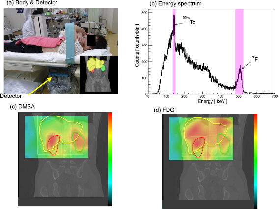
A Compton camera was placed under the right side of the volunteer's body to simultaneously detect RIs from the coronal views of the liver and kidney (figure 2(a)). Because the field of view was limited to the abdominal region by a lead shield, the bladder and most of the heart were not included in the imaging area. The estimated distance between the kidney and the Compton camera surface was 300 mm.
Figure 2. (a) Position setup with 2nd volunter (1st volunter's result is shown in supplemental figures). (b) Acquired energy spectrum through this study. (c) The accumulation 99mTc-DMSA, represented using a color contour map. (d) The accumulation of 18F-FDG, represented using a color contour map.
Download figure:
Standard image High-resolution image
On the first day of the human study, computed tomography (CT) images around the liver and kidneys were acquired. Reference positions were marked on the subject's body for comparison of the CT images with the Compton images. Then, on the second day, the volunteer was simultaneously injected with 99mTc-DMSA (30 MBq, Nihon Medi+Physics) and 18F-FDG (150 MBq, Nihon Medi+Physics); thereafter, four cyclic sequences involving 45 min of data acquisition and 15 min of rest were performed.
2.4. Image reconstruction method
List mode data consisting of channel IDs, pulse heights, and trigger timing, among other parameters, were recorded by the Compton camera. Based on the event selection method discussed in a previous study (Watanabe et al 2014), full-energy deposited Compton events were recorded within the energy window (e.g. 135–150 keV for 99mTc and 480–525 keV for 18F). In particular, the Compton events are back-projected onto an image plane parallel to the camera surface using a near-field back projection technique based on the method described in a previous study (Takeda et al 2012) with the value of the weighting parameter w set to 0. This projection is used to estimate the source flux at the projected plane based on the changes in flux due to the different distances of the detector from each point on the Compton cone. It should be noted that no likelihood estimation or deconvolution algorithms were included in the current study.
3. Results
The energy spectrum was processed from the detected events over a 35 min period from the 54 min to 89 min marks after RI injection; the spectrum is shown in figure 2(b). As can be seen from the figure, the energy peaks from both 99mTc-DMSA (141 keV) and 18F-FDG (511 keV) were clearly detected. Using the methods described in the previous section, the full-energy deposited Compton events were captured within the energy window (hatched region), and a total of 6741 and 4895 Compton events were extracted for 99mTc-DMSA and 18F-FDG, respectively. The events were back-projected onto the image plane placed 300 mm away from the camera surface, where the liver and kidneys were located.
The accumulation of 99mTc-DMSA is represented using a color contour map in figure 2(c); this map is overlapped by a reference CT image. As can be observed from the figure, two strong concentration areas of 99mTc-DMSA were clearly detected. Based on the reference CT images, these concentrations seemed to appear at the left and right kidneys. This assessment is supported by the well-known observation that 99mTc-DMSA tends to accumulate in healthy kidneys (Lin et al 2000, Sheehy et al 2009). In addition, we confirmed this result using a human abdomen phantom with plastic right and left kidneys filled with liquid 99mTc-DMSA with a radioactivity of 12 MBq; two intense concentrations of the isotope originating from the phantom kidneys were also clearly made visible, which firmly corroborated the abovementioned result. Conversely, 18F-FDG (figure 2(d)) was observed to be broadly distributed over regions that indicated the liver and kidneys, depicting a similar morphological structure when compared with the imaging results of 18F-FDG using PET-CT in routine clinical practice (Cheng et al 2013, Heusch et al 2013). Thus, the differences in kinetic and static behaviors inside the human body between the two radiopharmaceuticals 99mTc-DMSA and 18F-FDG were confirmed using Compton camera imaging.
4. Discussion
The objective of this study was to investigate the potential of a Si/CdTe Compton camera for simultaneous imaging of two radiopharmaceuticals (99mTc-DMSA and 18F-FDG) in the human body. Via our imaging experiments, we successfully demonstrated that the Compton camera was able to capture images of the distributions of 99mTc-DMSA and 18F-FDG, using specific 141 keV (99mTc) and 511 keV (18F) gamma rays in one exposure.
In this study, we conducted human imaging using 99mTc-DMSA and 18F-FDG. It is clear that other radioactive tracers attached to the same RI can be imaged by our Compton camera. In addition, the energy of 99mTc is low, whereas 18F generates higher energy gamma rays than a SPECT agent. Thus, the ability of simultaneous imaging to use 99mTc and 18F indicates that any SPECT tracers emitting 141–511 keV gamma rays (i.e. 123I, 111In) can be imaged by our Compton camera.
The gamma rays from both 99mTc and 18F were scattered in the body. The reduction ratio of the 141 keV gamma ray was estimated at 80%, considering the attenuation coefficient and the body thickness. In addition, some scattered gamma rays from 18F may have contaminated the data of 99mTc. However, our Compton camera was able to clearly image the kidneys because the cross-talk effect on the Compton imaging is relatively small in comparison to SPECT (Sakai et al 2018). This capability results from the Compton camera having excellent energy resolution, so the peaks of 99mTc and back-scattered gamma rays of 18F could be distinguished.
The contour of the liver was unfortunately indistinct in the image of 18F-FDG, in contrast to the contour of kidneys in the image of 99mTc-DMSA. This is because the 18F-FDG was distributed in other organs due to the absence of fasting, so spatial and contrast resolution of Compton camera was not sufficient. Though the imaging resolution of the prototype Si/CdTe Compton camera used in this study is not sufficient for clinical use and does not surpass that of SPECT and PET, the results of our study indicate high potential for the application of Compton cameras in the field of multi-energy RI tomography in the future.
The spatial resolution of a Compton camera is proportional to the distance from the detector to the source. In this study, the distance was 300 mm, but that distance can be reduced. Moving the Compton camera closer to the patient would improve the spatial resolution.
Compared with conventional SPECT, which suffers from count loss due to absorption in the collimator, overall efficiencies of Compton cameras are expected to improve in the future, enabling relatively shorter medical examination times than when SPECT is used.
The dosages of RIs administered in this study (30 MBq 99mTc-DMSA and 150 MBq 18F-FDG), which were sufficient to obtain clinically useful human images in 35 min, are already below the regulatory limits. Furthermore, a Si/CdTe Compton camera recently developed for applications in space (Schelbert et al 2002) has significantly improved efficiency. Thus, in the near future, it may surpass that of clinical SPECT. In a typical SPECT diagnosis, the injected dose of 99mTc-DMSA is 74–222 MBq (Veenboer et al 2015, Blaufox et al 2018), the efficiency is 0.01% to 0.1% (Van Audenhaege et al 2015, Slomka et al 2015), spatial resolution is approximately 10 mm (Van Audenhaege et al 2015, Slomka et al 2015), and the acquired time is 3–20 min (Maryse et al 2017, Freeman et al 2018, Fujiwara et al 2019).
Hence, the use of an advanced Compton camera as described herein has the potential to reduce human exposure to gamma rays from RIs during nuclear diagnosis in the future.
In this study, one Compton camera was placed underneath the body. Thus, the captured information was limited to a single angle of projection; therefore, the images obtained were limited to a 2D space. We have already successfully reconstructed the 2D and 3D distributions of different RIs in a rat by moving the camera around the rat to gather multi-angle data (Suzuki et al 2013, Sakai et al 2018). In the future, we will apply this monitoring technique to pre-clinical and clinical imaging in human.
In addition to its capacity for multi-probe tracking, we confirmed that the Si/CdTe Compton camera has other various advantageous characteristics, such as the potential to reduce examination time, a wide energy band, high energy resolution, and portability. In conclusion, our results indicate that the Si/CdTe Compton camera has great potential for various clinical applications in the future, and may facilitate new nuclear diagnostic procedures. Further improvements in its detection efficiency, spatial resolution, and image reconstruction algorithms are ongoing.
Acknowledgments
This work was supported by Grants-in-Aid from the Program for Leading Graduate Schools from the Japan Society for the Promotion of Science and for Scientific Research from the Ministry of Education, Culture, Sports, Science and Technology of Japan. The authors thank Dr Aiko Yamaguchi, Madoka Fujisaki, Dr Daisuke Irie, and Takayoshi Ishii for their assistance with the preparation of the manuscript. The authors have confirmed that any identifiable participants in this study have given their consent for publication.