


Abstract
An ultrathin layer of uranium nitrides (UN) has been coated on the inner surface of depleted uranium hohlraum (DUH), which has been proven by our experiment to prevent the oxidization of uranium (U) effectively. Comparative experiments between the novel depleted uranium hohlraum and pure golden (Au) hohlraum are implemented on an SGIII-prototype laser facility. Under a laser intensity of 6 × 10¹⁴ W cm−2, we observe that the hard x-ray (hν keV) fraction of the uranium hohlraum decreases by 61% and the peak intensity of the total x-ray flux (0.1 keV∼5.0 keV) increases by 5%. Radiation hydrodynamic code LARED is used to interpret the above observations. Our result for the first time indicates the advantages of the UN-coated DUH in generating a uniform x-ray source with a quasi-Planckian spectrum, which should have important applications in high energy density physics.
Export citation and abstract BibTeX RIS

Content from this work may be used under the terms of the Creative Commons Attribution 3.0 licence. Any further distribution of this work must maintain attribution to the author(s) and the title of the work, journal citation and DOI.
1. Introduction
In a hohlraum radiation source, powerful laser beams illuminate the high-Z inner wall of the hohlraum and then convert into intense x-rays from multi-eV to multi-keV [1, 2]. This x-ray source is initially designed to be the driven field for the indirect-drive inertial confinement fusion (ICF) [3]. Up to now the hohlraum radiation source has been applied in many other experiments, including studies on the high-pressure equation of state (EOS) of materials, explorations on opacity and radiation transport in astrophysics, and other research in high energy density physics [4, 5].
The most important factors for hohlraum radiation sources include the hohlraum radiation temperature (Tr), the x-ray spectrum and the spatial intensity distribution of the x-ray flux. In previous work, Tr has been successfully increased by adjusting laser parameters, hohlraum geometries and wall materials [5–7]. However, a homogeneous x-ray field with a Planckian or quasi-Planckian spectrum, which is highly desirable for many applications, still cannot be obtained by recent hohlraums. This problem is caused by the multi-keV hard x-rays, which usually make up a significant fraction of the total x-rays and result in a corresponding spectrum made of a superposition of a Planckian spectrum and a harder x-ray Gaussian spectrum [8]. In addition, most hard x-rays are emitted from the coronal plasma near the laser spots, so the flux intensity of hard x-rays is strongly anisotropic in space [9]. These problems are undesirable for most applications. In indirect-drive ICF where x-rays ablate the fusion capsule and finally ignite it, hard x-rays above 1.8 keV can penetrate beyond the ablation surface of ignition capsule, preheat the fusion fuel prior to the arrival of the shock front and then seriously degrade the implosion efficiency [10, 11]. To prevent the preheating effect, multi-layer doped ablators have been applied in ignition capsules [12]. However, adding dopant is a challenging technique that brings in large uncertainties. In some cases, the uncertainties might lead to serious hydrodynamic instabilities of the ablator and increase the mixing between the fuel and the ablator [13].
The ideal hohlraum radiation source for many applications should have both a high radiation temperature and a low hard x-ray fraction. In traditional cylindrical Au hohlraums, hard x-rays are usually more than 20% of the total x-rays [14, 15], because the hard x-rays are mostly generated by Au M-band emissions around 2.5 keV that are easily excited under a high radiation temperature. Dog-bone Au hohlraums can reduce the hard x-ray fraction by using a special structure to hide the M-band x-ray emitting region [16]. However, the dog bone structure also limits the radiation temperature. A depleted uranium hohlraum is considered as a better choice than a Au hohlraum. There are two main reasons: depleted uranium has a higher albedo (the ratio of thermal radiation emitted from the wall to that absorbed by the wall), meaning higher Tr, and U M-band emission around 4.0 keV is hard to excite, resulting in a low hard x-ray fraction [17]. Nevertheless uranium is very active and can be oxidized by the residual oxygen even in a high vacuum. The oxide layer of uranium is corrosive and will induce internal stress in bulk uranium, giving rise to cracks, crinkles and powders on the hohlraum interior. Cocktail hohlraum, which is made from a block of uranium coated with high-Z mixtures, theoretically has a higher opacity than either single material [18]. However, cocktail hohlraum has not been widely used due to the wall oxidization. The sandwich hohlraum recently applied in the National Ignition Campaign (NIC) has a sandwiched geometry of Au+U+Au [19]. The inner Au coating layer, which is 600 nm∼700 nm thick, can protect the middle U layer against oxidization [15, 20]. But the Au coating layer with a thickness of several hundred nanometers can emit intense M-band x-rays, counteracting the advantage of the middle U layer in hard x-ray emission. As a result, the hard x-ray fraction of the Au+U+Au hohlraum exceeds 14% when the radiation temperature is about 300 eV [15].
In this letter, we report a novel depleted uranium hohlraum (DUH), which preserves the sandwiched structure while replacing the inner Au coating with an ultrathin layer of depleted uranium nitride (UN). We have proven experimentally that the UN can prevent the oxidization of DUH effectively. Under a laser intensity of 6 × 10¹⁴ W cm−2, we observe that the hard x-ray fraction above 1.8 keV of U hohlraum decreases by 61% and the peak intensity of total x-ray flux (0.1 keV∼5.0 keV) increases by 5%, compared to Au hohlraum. We also extend our discussion to the hard x-ray emissivity of UN-coated DUH under ignition status and the influence of the reduced hard x-ray fraction on the implosion asymmetry.
2. UN-coated DUH against oxidation
We choose uranium nitrides as the coating material because UN has the potential to prevent the oxidation of uranium hohlraums. According to recently reported work [21], UN reacts with O ions to form UO2 and U2N3 after long-term storage in air. Also, the corrosivity of the final UN oxides is very slight compared to that of the U oxides. We sputter a UN layer of 100 nm thickness on the inner surface of a cylindrical depleted uranium hohlraum. The thickness fluctuation of the UN coating is controlled below 10 nm. Each newly fabricated UN-coated DUH is exposed to the air environment in the laboratory to test its oxidation endurance. Figures 1(a) and (b) show optical micrographs of uranium half-hohlraums with and without UN coating. Visible cracks and crinkles quickly appear in the surface of the uranium target without UN coating. In contrast, the UN-coated DUH has a smooth and clean inner surface. We further use x-ray photoelectron spectroscopy (XPS) to analyze the wall composition of a UN-coated DUH after an exposure test of 72 hours [22]. Figure 1(c) shows how the atomic percentages of oxygen, nitrogen and uranium in the wall material change with the depth from the inner wall surface. The oxygen content drops rapidly from 56.5% at the surface to 6.7% at about 3 nm below the surface, due to the free-state oxygen atoms adsorbed on the UN coating and the oxides of the UN surface. From the depth of 3 nm to the depth of 75 nm, the molar ratio of uranium and nitrogen is around 1:1, while the average oxygen content is kept below 1%, verifying the effective prevention of oxygen diffusion and uranium oxidation by the UN coating. Throughout the duration of our comparative experiment, no cracks or crinkles were observed on the inner wall of the UN-coated DUH, even under a more complicated working environment including mechanical vibration. All the results demonstrate that UN-coated DUH is fairly reliable as an x-ray radiation source.

Figure 1. The inner surface micrographs of the uranium half-hohlraum with (a) and without (b) UN coating. (c) Atomic percentage evolutions with the depth from inner wall surface for the major elements of UN-coated DUH acquired by XPS.
Download figure:
Standard image High-resolution image3. Experimental setup
The Rosseland mean opacity of UN is analogous to that of uranium, and nitrogen has almost no hard x-ray emission. We thus expect the influence of the UN layer on the x-ray field produced by DUH to be much smaller than that of a Au layer with the same thickness. Therefore, the UN-coated DUH is able to produce an x-ray field with a lower hard x-ray fraction and higher radiation temperature. To check the above idea, we carried out the comparative experiments on the SGIII-prototype laser facility using both UN-coated DUH and pure Au hohlraums. Figure 2 shows the experimental setup. The diameter and length of the empty hohlraum are both 1000 μm, and the diameter of the laser entrance hole (LEH) is 800 μm. The wall of UN-coated DUH consists of three layers: 100 nm-thick UN as the coating layer, 2 μm-thick depleted uranium in the middle and 22.9 μm-thick Au as the outer layer. The wall of the pure Au hohlraum is 25 μm-thick Au. Eight frequency-tripled laser beams (λ = 0.35 μm) inject into the hohlraum with an incident angle of 45◦ with respect to the hohlraum axis. All the lasers are 1 ns flattop pulse, and the total laser energy is 4.9 kJ. The diameter of the laser focal spot is adjusted to 500 μm by using the continuous phase plates. As shown in figure 2(a), the laser spots are initially located in the middle of the hohlraum with neighboring spots overlapped to form a closed loop. The laser intensity in the overlapping area reaches 6 × 10¹⁴ W cm−2. A transmission grating spectrometer (TGS) is installed at 20◦ to acquire the spectrum from 0.2 keV to 5.0 keV [23]. The TGS consists of one 2000 l/mm transmission grating and one detecting charge-coupled device (CCD). The diffraction efficiency of the transmission grating and the quantum efficiency of the CCD are absolutely calibrated on the Beijing Synchrotron Radiation Facility. The spatial and spectral resolution of TGS are 110 μm and 0.075 nm, respectively. Three groups of x-ray detectors (XRD) are set at 20◦, 30◦ and 45◦. Each group comprises two types of detectors, all of which are calibrated absolutely. A flat-response XRD (FXRD) measures the intensity of the total x-ray flux from 0.1 keV to 5.0 keV, through which the radiation temperature inside hohlraums can be obtained [24]. An M-band flat-response XRD measures the intensity of M-band x-ray flux from 1.6 keV to 4.4 keV [25]. An artificial spectrum composed of a Planckian distribution and a Gaussian distribution is involved to calculate the intensity of x-ray flux above 1.8 keV. The time resolution and intensity uncertainty of XRD are 100 ps and 10%, respectively. Two x-ray pinhole cameras are employed to confirm that the beams enter the LEH without striking the outside walls of the hohlraum and observe the LEH closure.

Figure 2. Schematic of experimental setup (a) and views of detectors (b).The red and yellow laser spots are produced by the top and bottom laser beams, respectively.
Download figure:
Standard image High-resolution image4. Results and analysis
In the comparative experiment, the total laser energy fluctuation is 3.8% and the maximum deviation of one laser beam's energy is 6.5%. The root mean square of the laser spot position offset is below 30 μm, and the maximum time offset of different laser beams in one shot is below 20 ps. As a consequence, the critical data including the x-ray fluxes and spectrums have good repeatability.
Figure 3 shows the radiation temperatures measured by FXRD and simulated by LARED (a two-dimensional radiation hydrodynamic code [26]). The measured peak Tr of Au hohlraums by detectors set at 20◦, 30◦ and 45◦ is 204 eV, 218 eV and 214 eV respectively, while that of UN-coated DUH is 207 eV, 220 eV and 218 eV correspondingly. The error bar of measured Tr is 2.5%, which includes the minor effect of the slight LEH closure during 1 ns. The Tr of 20◦ is lower than that at two other angles, due to the smallest fraction of the laser spot area in the view area at 20◦ as shown in figure 2(b). In these comparative experiments, we did not use the full aperture backscatter station or the near aperture backscatter station to measure the scattered light. In the simulation, the scattered light energy of Au hohlraum is set as 8% of the total laser energy. This value was obtained from previous Au hohlraum energetic experiments on SGIII-prototype [27], in which the total laser energy and Tr are similar to those of our comparative experiment. As the scattered laser energy of UN-coated DUH has not been measured yet, we assume it to be 8%, considering the same laser and similar plasma conditions as Au hohlraum. In order to simulate the Tr, the code also brings in U/Au gas with an initial density of 0.008 g cm−3 to fill the hohlraum, which does not exist in experiments. The faster rise of simulated Tr than measured after 0.75 ns could be ascribed to the x-ray emission of U/Au gas accumulating to the hohlraum axis. In spite of this, the simulated Tr agrees well with the experimental results in term of both temporal behavior and angular distribution. The increased temperature demonstrates that UN-coated depleted uranium has slightly higher laser to x-ray conversion efficiency than Au.
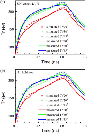
Figure 3. Experimental (solid lines) and simulated (stars) radiation temperatures of (a) UN-coated DUH and (b) Au hohlraum.
Download figure:
Standard image High-resolution imageFigure 4(a) shows the time-integrated x-ray images captured by the CCD of the transmission grating spectrometer, showing that the hard x-rays are significantly reduced in UN-coated DUH. The 900∼950 pixels of the CCD can observe the laser spot area. Compared with the Au hohlraum that gives about 30 000 counts around 2.5 keV, the UN-coated DUH exhibits almost no hard x-ray emission in the image and the CCD counts are only 3000. Meanwhile, the soft x-ray emission from UN-coated DUH has a distinct enhancement, which is consistent with the increasing trend of the radiation temperature. Figure 4(b) shows the unfolded spectra from figure 4(a). The M-band spectral shape of Au hohlraum has a double-peak structure, which is the same as the reported structure measured by a high-resolution crystal spectrometer [8]. The peak values are located at 2.1 keV and 2.6 keV, corresponding to the spectral line groups of 4p-3d and 4f-3d respectively. In the spectrum of UN-coated DUH, the N-band x-ray emission of UN-coated DUH blue-shifts from 0.8 keV to 1.2 keV, and the U M-band x-ray around 4.0 keV has not been emitted. As a result, the spectral shape of hard x-rays of UN-coated DUH is similar to that of a Planckian distribution. The integrated value of x-rays above 1.8 keV from UN-coated DUH is 65% lower than that from Au hohlraum.

Figure 4. (a) X-ray images captured by CCD of TGS, and (b) spectra of UN-coated DUH and Au hohlraum derived from the CCD images normalized by the peak value of the M-band spectrum of Au hohlraum.
Download figure:
Standard image High-resolution imageFigure 5 shows the temporal behavior of the spectrally integrated x-ray flux measured by the XRD group at 20◦. It is found that the UN-coated DUH produces 5% higher peak intensity of total x-ray flux than Au hohlraums. The integrated value of the hard x-ray flux of UN-coated DUH is 54% lower than that of Au hohlraums. The descending range of 54% measured by XRD is smaller than the 65% by TGS, because XRD determines an average intensity in the entire field of view whereas the detection area of TGS is only a small part of the XRD view area. The hard x-ray fraction is 12% for Au hohlraums and 4.7% for UN-coated DUH, which is the ratio of the integrated value of hard x rays to that of total x rays. The hard x-ray fraction of UN-coated DUH apparently decreases by 61%. In a Planckian radiation source with a Tr of 207 eV, the hard x-ray fraction is 2.5%, which indicates that the UN-coated DUH is a quasi-Planckian radiation source. Under the same initial hohlraum size and laser conditions, the Au plasma moves faster than the U plasma due to lower atomic mass, which narrows the hohlraum radius and increases the subsequent laser intensity. Meanwhile the faster movement of Au plasma also enhances the closure of LEH. The two factors counteract each other, resulting in a negligible influence on the x-ray flux measured outside the LEH. It is observed that the rising and falling rates of the total flux from UN-coated DUH are faster than those from Au hohlraums. A possible explanation is that the atomic processes of uranium are faster than those of gold, as the uranium atom has more outer electrons and its minimum ionization energy is less. The faster response of the x-ray flux to a laser pulse of UN-coated DUH is beneficial for shock shaping in ICF.

Figure 5. Total (0.1 keV∼5.0 keV) and hard x-ray (hν keV) fluxes of UN-coated DUH (solid) and Au hohlraums (hollow). The black circle curves correspond to the left-hand scale and the blue triangle curves correspond to the right-hand scale.
Download figure:
Standard image High-resolution image5. Discussion and conclusions
The above experimental results demonstrate that a UN-coated depleted uranium hohlraum can produce an x-ray source with ultra-low hard x-ray fraction and high radiation temperature, under the laser intensity of the SGIII-prototype laser facility. We then used LARED to simulate the spectrum under the ignition conditions. In the simulation the diameter and length of an empty hohlraum are 5.4 mm and 9.72 mm respectively, the diameter of the laser entrance hole (LEH) is 3.24 mm and the total laser energy is 1.45 MJ in 21.3 ns. The simulated peak Tr of the Au hohlraum and UN-coated DUH are 300 eV and 310 eV respectively. Figure 6(a) shows the simulated spectrum when the laser pulse is over. The U M-band spectrum around 4.0 keV is emitted and the hard x-ray part of the Planckian distribution is also enhanced. We subtract the Planckian part above 1.8 keV from the simulated spectrum in the same energy range, and then calculate the ratio of the remaining spectrum to the total x-ray spectrum. The ratio for the Au hohlraum is 0.04, while the ratio for UN-coated DUH is 0.02. Because the average atomic physics model in LARED is not precise, we also used a DCA code with a nonlocal thermodynamic equilibrium model [28] to calculate the hard x-ray emissivities of UN-coated depleted uranium and Au under ignition conditions. The simulation shows that both the peak and spectral-integrated hard x-ray emissivities of UN-coated uranium are much lower than those of gold when the electron temperature is up to 3.5 keV and the plasma density is 0.02 g cm−3, as shown in figure 6(b). This electron temperature corresponds to a hohlraum radiation field with a Tr above 300 eV. Both simulated results indicate that the advantage of UN-coated DUH in low hard x-ray emission can extend to the ignition status.

Figure 6. (a) Spectra of Au hohlraum and UN-coated DUH simulated by LARED. (b) X-ray emissivities of UN-coated uranium (thick blue line) and Au (thin red line) simulated by DCA code.
Download figure:
Standard image High-resolution image
The significantly decreased hard x-ray fraction upgrades the capability of hohlraum radiation source and can avoid some issues encountered in recent applications. In ICF, we predict that using UN-coated DUH will weaken the preheating effect and reduce the requirement to add dopants in capsules compared to other hohlraums under the same hohlraum geometry and laser conditions. In addition, the lower hard x-ray fraction leads to a more homogeneous intensity distribution of x-rays in space, which is expected to improve the implosion symmetry. We use the IRAD3D code [29] to interpret the influence of the decrease in hard x-rays on the asymmetry of the capsule. This code is based on analysis of the view factor, in which hard x-rays only distribute in the laser spot areas while soft x-rays distribute in both spot areas and re-emission areas with a weight ratio of 2:1 according to previous observations [9]. In a simple case in which eight laser beams inject into a cylindrical hohlraum to form two loops of laser spots, the calculated P2 asymmetry of the capsule by x-rays drops from 7.27% to 3.15%, if the hard x-ray fraction changes from 12% to 4.7%.
Based on the benefits in terms of preheating and asymmetry, we believe UN-coated depleted uranium is a promising material for the ignition hohlruam. Based on our previous study of the sandwich hohlraum wall of 0.1 μm Au + 5.0 μm U + 19.9 μm Au [19], we designed a novel wall composition of 0.1 μm UN + 5.0 μm U + 19.9 μm Au. Compared to the sandwich hohlruam of Au+U+Au applied in NIC, the UN+U+Au sandwich hohlraum can generate nearly the same radiation temperature but a lower hard x-ray fraction. The UN thickness of 100 nm is also enough to protect the DU layer of the ignition hohlraum against oxidation. However, regarding the much larger size of the ignition hohlraum than the hohlraum used in the comparative experiment, we expect fabricating such a large, thin, uniform UN coating layer uniform to be more difficult.
In summary, we propose that uranium nitrides as a coating layer can effectively prevent the oxidization of depleted uranium hohlraums and demonstrate the optimization on the x-ray spectrum and spatial uniformity by UN-coated DUH for the first time. We expect the novel hohlraum to be a better radiation source than a pure Au hohlraum or even a Au+U+Au sandwich hohlraum. Further work on fabricating gas-filled uranium hohlraums coated by UN and studying the improvements of implosion on the nearly completed SGIII laser facility with total laser energy of about 100 kJ in shaped pulses are currently under way.
Acknowledgments
This work was supported by the National Natural Science Foundation of China under grant no. 11475154.