

Abstract
Graphene plasmonics, with dynamic tunable resonance wavelength, has been successfully used in broadband plasmon-enhanced infrared spectroscopy. However, the requirement for external voltage loading makes the practical application sophisticated. In this work, the hybrid structure of graphene nanodot array (GNA) and gold nanoparticles (AuNPs) has been proposed as a passive platform for broadband infrared absorption enhancement. Numerical simulations show that the plasmon resonance peak of GNA becomes stiffer and broader when introducing AuNPs, and this is also proved by experimental results that the vibrational modes of polyethylene oxide molecule in a broad spectral range can be simultaneously enhanced. The metal-graphene hybrid plasmonic system has been proved to be a promising candidate for infrared sensing, which is significant for safety and healthy applications.
Export citation and abstract BibTeX RIS
1. Introduction
Graphene, as a promising alternative plasmonic material beyond conventional noble metals, offers the potential of versatile photonics device applications due to its unique resonance tenability [1–4], high degree of electromagnetic confinement [5, 6] and presumably long plasmon lifetime [1, 5]. Indeed, graphene plasmon has already shown utility in enhancing infrared absorption for surface-based biological and chemical sensors [7–11]. Li et al firstly realized detection of thin layers of solid polymers through graphene plasmon enhanced infrared spectroscopy [7]. Then, Rodrigo et al demonstrated a high-sensitivity infrared absorption detection in an electrically tunable broad spectral range [9]. Simultaneously, Farmer et al detected vibrational modes in small quantities of gas-phase molecules using tunable graphene plasmons [10]. In a recent study, Hu et al designed a graphene-based infrared sensor that covers the entire molecular fingerprint region by utilizing the tunable graphene plasmon platform [11]. However, the above dynamic tuning approaches require external voltage loading, which makes the practical application sophisticated. On the other hand, it has been demonstrated that plasmon resonances and associated optical fields in graphene can be tuned by modifying the carrier density [12], the dielectric constant of the surrounding media [13], and the critical size of the micro-/nano-patterning arrays [14–17]. Therefore, metal nanoparticles could be an excellent choice to be introduced into graphene platform. The advantages are two fold: they are not only additionally employed as infrared absorption enhancer [18], but also changing the electrostatic environment around graphene [19]. Moreover, metal nanoparticles could be good doping source for graphene [20, 21], which will result in stronger resonances to offer larger near-field enhancement and stronger infrared absorption [22].
In this letter, we propose a hybrid structure, consisting of graphene nanodot array (GNA) and gold nanoparticles (AuNPs), to enhance the infrared absorption of analytes. The hybrid structure is fabricated by a readily approach of block copolymer self-assembly and a rapid thermal annealing of gold film. Through measuring infrared transmission, we found an enhancement of absorption of incident light for the proposed hybrid structures compared with bare GNA. In comparison with previous works, our results facilitate a better understanding of the plasmon coupling between GNA and AuNPs. In addition, we demonstrate the broadband detection of vibrational modes of polyethylene oxide (PEO) molecule in submonolayer thickness. We believe our work is promising to offer superior detection capabilities compared with conventional plasmonic sensors.
2. Experimental details
2.1. Block copolymer nanopatterning of GNA
The graphene for the GNA was grown on copper (Cu) foil by low-pressure chemical vapor deposition and transferred on a highly resistive, double-side polished Si substrate using a common wet transfer method. Then, the graphene on the substrate was immersed in a dilute aqueous solution of dopamine (1 mg ml−1), buffered to a pH typical of marine environments (10 mM Tris-HCL, pH = 8.0) for 1 h followed by rinsing in deionised water. After that, a thin film of polystyrene-block-poly(2-vinylpyridine) (PS-b-P2VP) (5700-b-5700 g mol−1) reverse micells was coated from m-xylene solutions at a polymer concentration of 0.5% (w/w) on to the graphene by spin-coating at 5000 rpm. Patterning of the underlying graphene was carried out by the O2 plasma (100 W, 20 Pa, 20 sccm, 65 s). Finally, the samples is annealed by vacuum high-temperature furnace (1e−4 Pa, 700 °C, 2 h) to remove protection polymer layer of graphene nanodots.
2.2. Preparation of the AuNPs-GNA hybrid structure
The 10 nm thick gold film was deposited on the graphene nanodots/Si substrate at a rate of about 1 nm s−1 and at a power of ∼200 W by magnetron sputtering. Then, the gold film was subjected to a rapid thermal annealing step at 400 °C for 5 min in N2 atmosphere to obtain the AuNPs.
2.3. PEO molecule for plasmonic sensing
A 0.06% (w/w) PEO polymer dissolved in methyl alcohol was dropped on the samples for sensing detection. The 5% (w/w) PEO polymer is used for the reference concentration.
2.4. Characterizations
The AuNPs–GNA hybrid structure were characterized by scanning electron microscopy (Hitachi S-4800). The surface morphology and thickness of the samples were studied by atomic force microscope (Bruker Dimension ICON). The Raman spectra were obtained by Raman microscopy (Horiba Jobin Yvon LabRAM HR800) with a 532 nm excitation laser. X-ray photoelectron spectroscopy (XPS) measurements were performed on an EscaLab 250Xi instrument with a monochromated Al Kα source (1486.68 eV). The Fourier transform infrared spectroscopy (FTIR) extinction spectra were performed using a Bruker V80 vacuum interferometer equipped with a Globar infrared source and KBr beam splitter.
3. Results and discussion
The schematic of the hybrid structure is shown in figure 1(a), where AuNPs are deposited on GNA. The absorption spectra is acquired by transmission mode of (FTIR). The hybrid structure is prepared by a block-copolymer (BCP) self-assembly method that is versatile bottom-up approach capable of achieving graphene patterned arrays at the wafer-level and a rapid thermal annealing of gold film to obtain AuNPs, which has advantages of more cost-effective and a higher output than the traditional fabrication of GNA by electron beam lithography. After transferring and surface modification of graphene, polystyrene-b-poly(2-vinylpyridine) (PS-b-P2VP) is directly spin-cast on graphene, which performs as a nano-feature mask to pattern graphene. The P2VP block of the copolymer forms the core and is responsible for pattern formation while the PS block of the BCP forms a thin continuous layer. Figure 1(b) shows the patterned graphene template with a height of ∼17 nm and diameter of ∼100 nm. After achieving the patterned arrays of cylindrical microdomains with a high degree of lateral order, the BCP template is etched by O2 plasma for 65 s, resulting in patterning of the underlying graphene, i.e., GNA (the quality of graphene is shown in figure S1 is available online at stacks.iop.org/NANO/28/264001/mmedia in supporting information (SI)). Then, the sample undergoes annealing treatment to remove residual polymer. After the preparation of GNA, AuNPs are introduced (see figure 1(c)) by deposition and dewetting, the average diameter of AuNP is ∼66.91 nm. The XPS spectra clearly indicate the C 1s and Au 4f of the hybrid structure. Eventually, the AuNPs on GNA is shown in figure 1(d). More details are mentioned in the experimental sections.

Figure 1. (a) Mid-infrared transmittance measurement scheme for the AuNPs-GNA hybrid structure. (b) AFM image of as-spin coated PS-b-P2VP with self-assembled arrays. Inset: corresponding line-scan profile of b. (c) AFM image of the dewetted AuNPs on GNA. Inset: size distribution of AuNPs on GNA. (d) SEM image of AuNPs-GNA on Si substrate. The light region is covered by AuNPs. (e) C 1s and (f) Au 4f XPS of the hybrid structure.
Download figure:
Standard image High-resolution imageFinite-domain time-domain simulations were carried out to study the influence of a AuNP loading on the electric field of a graphene nanodot. As shown in figure 2(a), the larger area overlap between the AuNP and graphene nanodot, which indicates a stronger coupling, the bigger electric field enhancement factor at the edge of graphene nanodot can be obtained. It can be seen that the electric field intensity can be enhanced as large as three orders of magnitude at the GNA edge. Figure 2(b) shows the calculated extinction spectra, i.e. (1 − T/T0), of GNA with AuNP at different positions, where T and T0 are the optical transmission through the samples and the bare Si substrate, respectively. A clear resonance blue shift is observed for all cases. Besides, the graphene plasmonic resonance peak becomes significant broader, especially for highly overlapped situation.
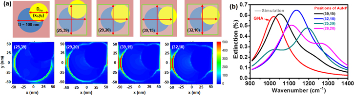
Figure 2. (a) The schematic of a gold nanoparticle on a graphene nanodot with diameter of 100 nm at different position and corresponding normalized electric field intensity distribution. The center coordinates of gold nanoparticle is relative to the center of graphene nanodot. (b) Calculated extinction spectra of bare GNA (the red curve) and AuNPs-GNA hybrid structure on Si substrate.
Download figure:
Standard image High-resolution imageBecause of annealing treatment process used in AuNPs preparation, the size of AuNPs distributed on the GNA is random. Figure 3(a) shows that the GNA are partly covered with the AuNPs in different sizes, and the corresponding normalized electric field intensity distribution is shown in figure 3(b). Compared with the bare GNA, the calculated extinction spectrum for the hybrid structure appears to be much broader as shown in figure 3(c). The extinction spectra of the prepared hybrid structure is measured by FTIR. As shown in figure 3(d), a prominent plasmonic resonance peak appears in the mid-infrared (MIR) region for bare GNA. The excitation of dipole resonance in bare GNA results in enhanced absorption, thus the spectral feature evolves into the strong plasmonic resonance around 1080 cm−1. On the other hand, the introduction of AuNPs results in an additional resonance peak at higher energies, which can convert incident light into graphene plasmons in a broader spectral range.

Figure 3. (a) 5 × 5 GNA unit partly covered by AuNPs, with size generated randomly in the range from 24 to 130 nm. (b) Normalized electric field intensity distribution of a. (c) Calculated extinction spectra of the 25 AuNPs randomly distributed on GNA. (d) Experimental extinction spectra of the prepared AuNPs-GNA hybrid structure.
Download figure:
Standard image High-resolution imageIn practical plasmon-enhanced infrared absorption application, we use PEO polymer, which possesses multiple resonance modes in the MIR range from 1200 to 1500 cm−1 [11] (see figure 4(a)), as the analyte to demonstrate the broadband enhancement ability of our hybrid structure. It is shown in figure 4(b) that the infrared absorption of sub-monolayer PEO (figure S2 in supporting information (SI)) on Si substrate is extremely weak (only mode B can be observed). After introducing GNA, the absorption intensity of mode D can be dramatically improved while no obvious enhancement for other modes can be detected, and this should be attributed to the narrow resonance peak of GNA. In contrast, great enhancement for the modes A, B, D, E, F has been observed with GNA-AuNPs hybrid structure and the enhancement factor for mode D is even larger than that of GNA, which indicates the higher electric field enhancement factor in a broader spectral range. The experimental results obtained here are in good agreement with the numerical simulations. Previous research on graphene plasmonic sensing has achieved ultrathin (8 m thickness) PEO film detection, while it is demonstrated in this work that the sensitive detection of sub-monolayer PEO molecules is available. The signal intensity of the mode A located at 1240 cm−1 shows ten-fold enhancement with our hybrid structure.

Figure 4. (a) The characteristic infrared absorption peaks of PEO molecule and the positions of corresponding vibrational modes; (b) bare GNA (blue line) and AuNPs-GNA hybrid structure (red line) enhanced infrared absorption of sub-monolayer PEO molecule.
Download figure:
Standard image High-resolution image
4. Conclusion
In summary, we have demonstrated the fabrication of GNA-AuNPs hybrid structure with the aid of the block copolymer self-assembly method and rapid thermal annealing process. The dipole resonance of the GNA appears in the MIR region. By introducing AuNPs, the plasmon resonance peak of GNA becomes stiffer and broader, which is ideal for broadband plasmon-enhanced infrared spectroscopy. The vibrational modes of PEO molecule in a broad spectral range has been successfully detected with the GNA-AuNPs hybrid structure, which is in good consistent with numerical predications. Therefore, the metal-graphene hybrid plasmonic system is a promising candidate for infrared sensing, which is practically meaningful for safety and healthy applications.
Acknowledgments
This work was supported by the Hundred Talent Program of Chinese Academy of Sciences, the National Natural Science Foundation of China (11574349) and the Natural Science Foundation of Jiangsu province (BK20150365). The financial support from the Opened Fund of the State Key Laboratory of Optoelectronic Materials and Technologies (Sun Yat-sen Unversity) is also acknowledged. ZP thanks the start-up Grant from Southwest University (No.SWU111010) for support. C-W Q acknowledges the A*STAR SERC Pharos grant No. 152700014 with project No. R-263-000-B91-305. LZ acknowledges the financial support from Young Talent Recruiting Plans of Xi'an Jiaotong University.