
Abstract
We report on the ionization and nanoplasma dynamics of small xenon clusters irradiated by intense, short pulses of a short-wavelength free-electron laser. Fluorescence spectroscopy indicates that inelastic electron collisions play a prominent role in the formation of the highest charge states. From the spectral distribution an electron temperature of 27 eV is deduced which corresponds to the average excess energy of the Auger- and photoelectrons ionized from individual atoms but trapped in the cluster core. This suggests that fluorescence spectra reflect a very early stage within the nanoplasma dynamics and shows how a part of the kinetic energy of the plasma electrons trapped in the cluster potential is transferred to the ions.
Export citation and abstract BibTeX RIS

Content from this work may be used under the terms of the Creative Commons Attribution 3.0 licence. Any further distribution of this work must maintain attribution to the author(s) and the title of the work, journal citation and DOI.
This article was made open access on 13 August 2015
1. Introduction
Nanoparticles exposed to intense and short laser pulses have opened an entirely new regime of matter–light interaction, leading to the formation of isolated, transient, solid density nanoplasmas [1, 2]. These transient states of matter can be observed at a wide excitation energy regime as shown by particle-laser interaction studies from the infrared (IR), towards the extreme ultraviolet (XUV) and x-ray regime provided by novel light sources such as higher-harmonic generation (HHG) systems and free-electron lasers (FEL) [3–14]. Nanoplasma formation is a direct consequence of strong irradiation. The formation and evolution of such plasmas are of fundamental interest to particle-FEL-interaction experiments, especially those involving imaging techniques [15–18].
During the last decade, rare gas clusters have proven themselves as model systems to study the nanoplasma induced by FEL pulses [1, 6–8, 16, 17, 19–34]. They can be tuned in size from few atoms [35] up to large diameters of more than one μm [36] which allows for studying size effects over a very large range. Further, their simple structure provides a direct insight into light-induced dynamics simplifying the analysis compared to complex chemical structures like e. g. biomolecules.
Upon FEL irradiation, clusters are initially ionized by photo-absorption until further electron emission is frustrated due to the increasing cluster Coulomb potential [21]. At higher power densities, frustration is reached during the pulse while electron removal from individual cluster atoms still proceeds (inner ionization [37]) and leads to nanoplasma formation [1, 27, 28]. In contrast to the IR wavelength range, field ionization and inverse bremsstrahlung, as well as simultaneous multiphoton ionization can be neglected in the XUV and x-ray regime for power densities below 1017 W cm−2 [1, 2]. For FEL wavelengths shorter than  nm, single direct photoionization dominates the energy deposition in the cluster [21, 28]. Therefore, the initial electron temperature of the nanoplasma is determined by the excess energy of the inner ionized photoelectrons and the subsequently emitted Auger electrons (termed ionization heating [38]). Depending on the electron density and temperature, the cluster subsequently disintegrates in a Coulomb explosion and/or a hydrodynamic expansion [28]. Large clusters are found to expand hydrodynamically accompanied by efficient recombination of the cluster ion core in the dense nanoplasma [22, 34, 39–41]. However, recombination is most probable when the nanoplasma is dense and cold. But therefore, it is necessary that the plasma electrons reduce their kinetic energy.
Theory predicts that electrons distribute their energy via collisional processes, and thereby also contribute to the inner ionization of the cluster [34, 42, 43]. So far, the role of electron impact ionization on the formation of charge states in FEL induced nanoplasmas has not been experimentally quantified. As fluorescence spectroscopy is sensitive to charge states as well as excitation and relaxation processes, it gives insight into electron impact driven processes. Furthermore, fluorescence is not affected by the large space charge of the cluster and is therefore more suitable to provide information on the role of inelastic electron collisions than ion and electron spectroscopy [44–46].
In this article we present a study of FEL-induced fluorescence emitted from small xenon clusters comprising an average amount of  up to
up to  atoms. We discuss the contributions of different ionization and excitation mechanisms and show that electron impact ionization and excitation is the dominant process for the formation of the high charge states. Since the excited states are mainly populated by electron collisions, fluorescence provides access to the electron temperature. We observe an electron temperature close to the electron excess energy, which suggests that the fluorescence reflects a very early state of the nanoplasma evolution when electron cooling has just started.
2. Experimental setup
The experiment was carried out in a tandem configuration at the free-electron laser in Hamburg (FLASH) at 90 eV photon energy with typical pulse durations of 150 fs and average pulse energies of around 100 μJ. In a back-reflecting geometry, a multilayer mirror refocused the FEL beam into a 2.5 μm spot (FWHM), yielding power densities exceeding 1015 W cm−2. Xenon clusters were produced in supersonic expansion through a conical nozzle (100 μm, 15° half opening angle). The cluster size distribution was controlled by the stagnation pressure and quantitatively estimated with the help of scaling laws [35]. To attain high target densities, the FEL focus was overlapped with the xenon cluster jet just in front of the nozzle. The radiation emitted from the FEL excited clusters was detected with a fluorescence spectrometer under an angle of 90° (minimal elastic light scattering). A toroidal grating (Horiba Jobin Yvon, type 541 00 200) focused and dispersed the fluorescence lines on a flat, XUV sensitive CCD camera. The fluorescence was recorded in the wavelength regime from 10 to 76 nm with a measured resolution of 0.2 nm [45]. The spectra are corrected for the grating efficiency and normalized according to the integration time and the target density, which scales in first approximation linearly with the stagnation pressure at the nozzle. For additional reference measurements an ion spectrometer could be included in the setup. Further experimental details and a scheme of the setup are published in [45].
3. Results and discussion
3.1. General aspects
In order to distinguish between atomic and cluster specific signatures we discuss fluorescence spectra from clusters in comparison to measurements on the atomic beam. Figure 1 displays the fluorescence from the atomic (black) and the cluster beam (red) as a function of wavelength. The average cluster size of the cluster beam was  . The atomic beam was generated by expanding gas at low pressures below the onset of condensation. In order to achieve an adequate signal to noise ratio, the integration time was increased for the atomic beam. Note that the fluorescence yield of the atomic spectrum is much smaller than its cluster counterpart and has been scaled with a factor of ten for better visualization. The signal from elastically scattered light of the FEL at 13.5 nm is marked blue. Xenon atoms as well as xenon clusters emit a multitude of fluorescence lines in the wavelength regime from 10–76 nm, associated to charge states from
  to
to  . The lines are identified with the aid of the NIST data base [47]. However, only very few reference data of charge states from
  to
to  as well as transitions from higher excited states to lower excited states for the charge states
  with
with  can be found. The fluorescence lines in the range from 10 to 20 nm, corresponding to high charge states, could be assigned by comparison to discharge measurements [48–50]. All in all, almost 80% of the altogether 120 lines could be assigned.
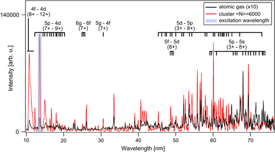
Figure 1. Comparison of fluorescence spectra from cluster (black) and atomic (red) beams, both acquired at an FEL power density of around 2·1015 W cm−2. The spectra are normalized to the same target density in the interaction volume and integration time. After normalization, the atomic spectrum is still much lower. Therefore, the intensity of the atomic spectrum is scaled with a factor of ten for better visibility. The highlighted peak (blue) is due to scattered light at the excitation wavelength of 13.5 nm. Spectral lines stemming from charge states between  and
and  are indicated for atoms as well as clusters.
Download figure:
Standard image High-resolution imageA quantitative analysis of the line intensities reveals a drastically increased total fluorescence yield from clusters in comparison to atoms. The total emission yield per atom calculated from the cluster spectrum, that is displayed in figure 1 is approximately a factor of 5 higher than the atomic fluorescence yield. Further, strong differences in the line intensity ratios are visible. This suggests that in clusters additional ionization and excitation processes take place.
In the atomic beam, photoionization, photoexcitation and intra-atomic relaxation are the only processes creating and exciting the ions. We like to note, that for charge states  with
with  , more than one single photon has to be absorbed in each ionization step [51]. Whether higher charge states are formed by sequential multiphoton absorption or collective electron excitation in the vicinity of the 4d giant resonance of xenon is still under discussion [51–56]. However, it is clear that in the atomic beam electron impact ionization and excitation are negligible because the target density is so low that the mean free path for the electrons is much longer than the diameter of the beam. Therefore, the comparison of the atomic spectrum with the cluster spectrum gives a direct measure of the contribution of photoabsorption and excitation to the total fluorescence yield. Obviously, the much stronger per-atom-fluorescence yield from clusters gives evidence that in the case of clusters mechanisms other than direct photon-induced processes contribute significantly. There are two additional possibilities to populate fluorescing states in clusters: electron impact and recombination related processes. Fluorescence allows to analyze the contribution of both mechanisms, which will be discussed in the following two sections.
3.2. The role of electron–ion collisions
The difference in fluorescence yield between cluster spectra and atomic spectra is most prominent for the highest charge states found in the wavelength range from 10 to 12 nm (cf figure 1). We would like to point out that these fluorescence lines are found at shorter wavelength than the excitation wavelength of 13.5 nm and belong, predominantly, to the 4f–4d transitions of the charge states from  up to
up to  , possibly even
  . A similar upconversion was already observed in the fluorescence spectra of argon clusters and Ar–Xe-core shell clusters [44–46].
Figure 2 displays the integrated 4f–4d fluorescence yield depending on (a) FEL power density and (b) cluster size. The yield of these fluorescence lines follows a step-like function with increasing FEL power density. Above the level of 5·1013 W cm−2 the signal immediately saturates. A size dependent investigation, shown in figure 2(b), reveals that the yield due to the 4f–4d transitions increases rapidly for small clusters, reaches a maximum at 300 atoms per cluster and decreases for larger cluster sizes.
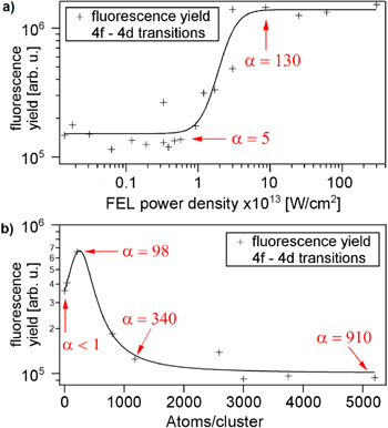
Figure 2. Total fluorescence yield of the 4f–4d transitions as a function of FEL power density at a cluster size of  atoms per cluster (a) and as a function of cluster size at an FEL power density of
  Wcm−2 (b). The line intensities of the 4f–4d transitions steeply rise as soon as the build-up of the positive cluster ion core traps the electrons and a nanoplasma evolves.
Download figure:
Standard image High-resolution imageWe attribute the sudden increase of signal with the FEL power density and the cluster size to the onset of nanoplasma formation, when electron emission becomes frustrated by the increasing Coulomb potential of the cluster. This denotes also the point where electron collision processes start to become efficient. This onset can be estimated through the frustration parameter  introduced by [28], which is the ratio of photo-activated electrons
  to outer ionized [37] electrons
  estimated for a spherical, homogenously charged cluster [28]. For
  , the cluster starts to trap a fraction of the photo-activated electrons leading to nanoplasma formation. Increasing α denotes that frustration sets in earlier and a nanoplasma is formed more efficiently. The parameter α is directly depending on FEL power density I0 as well as the amount of atoms per cluster N. In figure 2 the corresponding α values are calculated for the characteristic points of the graphs (indicated by the red arrows). The α values are calculated according to our experimental conditions: photon energy
  , pulse duration
  , photoionization cross section
  , Wigner–Seitz radius
  , ionization potential of the 4d shell
  .
.
In both cases, the steep rise of the yield of the 4f–4d fluorescence lines is in line with the transition of the frustration parameter from low to high values denoting that the cluster becomes able to efficiently trap electrons. As recombination rivals the fluorescence of the high charge states, it is natural that the fluorescence signal of the 4f–4d transitions of  to
to  decreases again with increasing cluster size (cf figure 2(b)), when recombination becomes efficient. The fast saturation of the 4f–4d transitions as a function of the FEL power density (see figure 2(a)) is presently not completely understood, and further investigations including theoretical work are necessary.
In order to obtain detailed insight into the role of inelastic electron collisions, we compare in the following the FEL-induced fluorescence from the highest charge states in our spectra to the fluorescence of xenon gas induced by electron impact [48]. Figure 3 is a small section of figure 1, displaying the fluorescence spectra in the wavelength range from 10 to 17 nm from clusters (red) and atomic gas (blue, x10). In addition, a spectrum produced by a dense plasma focus pinch discharge of xenon gas (black) from the literature [48] is added for comparison. The spectra are normalized to the same area under the peaks between 14 and 15.2 nm except the atomic spectrum. The line distribution in figure 3 shows a good match between the cluster fluorescence and the spectrum from the pinch discharge. Fluorescence from  ions overlappes with the elastically scattered light peak at 13.5 nm and therefore is not expected to agree with that of the pinch discharge spectrum. In XUV-excited clusters, compared to the purely collisionally excited atomic spectrum, a slightly higher yield for the group of lines between 10 and 11.8 nm is found. It can be explained by taking the contribution from photoabsorption processes observed in the atomic spectrum (blue) into account. The striking similarity between the cluster spectrum and the electron impact induced fluorescence spectrum (figure 3) as well as the rapid increase of the highest charge states with the onset of the nanoplasma formation (figure 2) convey a clear picture of inelastic electron collisions being the leading process for ionization and excitation in particular for the highest occurring charge states.
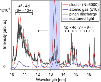
Figure 3. The FEL-induced fluorescence spectra of clusters (red) and atoms (blue) are compared with a fluorescence spectrum induced by a dense plasma focus pinch discharge (black). The fluorescence induced solely due to electron collisions in the pinch discharge matches all features of the cluster spectrum. The yield of the highest charge states formed in clusters is slightly above the fluorescence induced by electron collisions which might be an indication of photoabsorption at least contributing to some extend to the total fluorescence signal in the present experiment.
Download figure:
Standard image High-resolution imageFor understanding the efficient formation of high charge states via electron impacts several arguments can be made. In the cluster environment the ionization potentials of the cluster atoms and ions are lowered [57, 58]. Thereby collisional ionization of even highly charged ions becomes possible for electrons with lower kinetic energies than needed for isolated atoms. Both, photoionization and electron impact ionization, are enhanced by ionization potential lowering. Considering that every photoionization event releases at least one electron and taking into account the respective cross sections leads to much higher probabilities for collisional processes compared to photoabsorption.
Photoionization of xenon atoms from neutral xenon up to  at 90 eV photon energy is dominated by the 4d giant resonance with an absorption cross section of around 25 Mb [59]. The photoionization cross sections for higher charges, for the valence orbitals 5s and 5p, and for excited states, are more than an order of magnitude lower [59, 60]. Photoexcitation is even more unlikely because excitation is only possible if the photon energy matches dipol-allowed transitions to excited states. In contrast to the photoabsorption, an electron impact can transfer an arbitrary amount of kinetic energy. Electron impact ionization of xenon exhibits large cross sections for the valence shell. For example: at small kinetic energies of 22 eV, corresponding to the excess energy of a photoelectron from the 4d shell, the collisional ionization cross section exceeds already 140 Mb [61].
Recent modeling of XUV excited argon clusters indicates that also collisional two-step processes contribute significantly to ionization [43]. Cluster atoms are first excited and then ionized via inelastic electron collisions. This process is termed augmented collisional ionization [43]. It is found to occur much more frequently than direct electron impact ionization from the ground state, because each electron needs less kinetic energy. For xenon clusters initially ionized with 90 eV photons, this effect can be expected to play an even more important role since the photoelectrons in xenon have a slightly higher excess energy, and the excitation cross sections are much larger than in argon [62]. For example, the total collisional excitation cross section of neutral xenon for the ground state electrons has its maximum of 400 Mb at 11 eV kinetic energy [63]. For excited states, the electron impact ionization cross sections are even higher than in the ground state and can reach values up to 2500 Mb [62]. The cross sections for collisional processes at the relevant energies exceed the photoabsorbtion cross sections in every single excitation and ionization channel and lead to mean free path lengths around 1 Å which is more than an order of magnitude smaller than the cluster radius ( ). Along these lines we conclude that multistep collisional processes are the doorway to reach high charge states with low electron kinetic energies.
3.3. Influence of electron–ion recombination
Recombination can be a rivaling process and a contributor to fluorescence at the same time. If a plasma electron recombines with an Xe ion to
  it suppresses the fluorescence of the Xe
 species. But it is also very likely that the electrons recombine into long lived, weakly bound atomic Rydberg states [11, 57]. As an example a three-body recombination process is depicted in figure 4(a). When the electrons from the Rydberg states relax into lower-lying states afterwards, there is a probability for radiative decay which would be visible as a contribution to the fluorescence spectrum (see middle scheme of figure 4(a)). It is possible to investigate the contribution of recombination to the fluorescence yield through the application of a static electrical dc field across the interaction region. According to theoretical work, a weak electric field, such as the extraction voltage of a time-of-flight spectrometer (TOF), re-ionizes the recombined electrons from the weakly bound atomic Rydberg states [57]. This effect has recently been confirmed in experiments using an HHG source [11]. If recombination is suppressed as displayed in the lowest scheme of figure 4(a), the subsequent radiative decay of highly excited states cannot take place and some fluorescence lines should be diminished in the spectra.
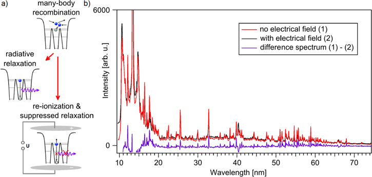
Figure 4. Influence of the dc extraction field of an ion spectrometer on the spectral fluorescence yield. Only small changes in the line intensity ratios are observed giving evidence that recombination contributes but is not the main process for the population of the fluorescing states.
Download figure:
Standard image High-resolution imageIn figure 4(b), two spectra recorded under comparable experimental conditions, are shown: one without (red) and one with (black) an applied external field of 1 kV cm−1. From the difference spectrum, we can estimate that the fluorescence stemming from recombination processes makes only a few percent of the total yield. It indicates that recombination related processes are of minor importance in populating the fluorescing states.
3.4. Electron temperature of the nanoplasma
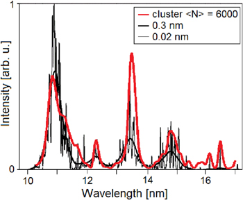
The electron temperature and the electron density of the nanoplasma determine the cluster nanoplasma evolution in this excitation wavelength range. Therefore, it is essential to have experimental access to these parameters. Provided that most of the fluorescence excitation is due to inelastic electron collisions with Xe ions, the electron temperature of the nanoplasma is imprinted in the spectral distribution. In the literature, the fluorescence for different average kinetic energies (electron temperatures) is calculated in the corona model and presented [48]. The calculations show, that slight changes of 1 or 2 eV of the electron temperature already lead to strong changes in the spectral line distribution. As shown in figure 5, the calculated spectrum for an average kinetic energy of 27 eV matches our fluorescence spectrum from xenon clusters.
Figure 5. Cluster fluorescence spectrum (red) and calculated fluorescence spectra for an electron temperature of 27 eV with resolutions of  and
and  . Adapted from [48].
Download figure:
Standard image High-resolution image
Even though the nanoplasma of the present experiment does not match the density of the calculated plasma we can use the calculations for estimating the electron temperature of the nanoplasma in clusters. The plasma density determines the yield for each single line but does not affect the intensity ratios. In the first place, the intensity ratios between the fluorescence lines depends only on the electron temperature. Nevertheless, other plasma characteristics like the opacity and the extent of the plasma can influence the measured spectral distribution via reabsorption and thereby distort the determined electron temperature. Due to a large extent but a low density of the calculated pinch discharge plasma a slight opacity factor (constant for all wavelength) has been taken into account within the calculations. In the present experiment the situation is reversed: the density is quite high but the extent of the nanoplasma is limited by the small cluster size of only a few nanometers. The line intensity reduction in our experiment is approximately a factor of 1.2 times stronger than in the calculations which might be the origin of the small deviations between the calculated spectrum and our measured spectrum.
The determined value of 27 eV coincides with the mean between the excess energy of a 4d photoelectron of 22 eV and the subsequent Auger electrons of 32 eV kinetic energy. It can be considered rather high, given that the electron temperature is expected to decrease quickly due to energy loss via inelastic collisions with ions, expansion and evaporation of hot electrons.
Thus, we assume that the fluorescing states of the highest charge states are populated in an early stage of the nanoplasma evolution. This conclusion is in line with our result that recombination is inefficient on the time scale of fluorescence. Beyond that, previous studies of heterogeneous clusters revealed fluorescence lines from high transient charge states, not observed in the final ion TOF charge state distribution. The earlier results imply, that such states are not only excited at the very beginning of the plasma dynamics but also can relax before recombination processes become dominant [45].
4. Summary and discussion
We have investigated the light-induced processes in small clusters with up to 6000 atoms, by means of fluorescence spectroscopy after excitation with intense XUV FEL pulses. We find evidence that the formation and excitation of the highest charge states in a cluster is governed by inelastic electron–ion collisions. Although the initial ionization of the cluster atoms is driven by photoabsorption, the results indicate that electrons already contribute significantly to the formation of charge states already from the beginning of the pulse and dominate as soon as the photoelectron emission of the cluster gets frustrated. Further, collisional processes provide a fast and efficient energy transfer mechanism in clusters. The electrons distribute most of their energy via inelastic collisions to the ions, either exciting or further ionizing them, while the kinetic energy of the ions stays almost the same.
Recombination has been found to be an important process later in the nanoplasma evolution [12, 22, 34, 40, 41, 60] when the electron temperature is low, typically after some hundred femtoseconds [34, 40]. The majority of fluorescing states is populated by inelastic collisions with energetic electrons. This means that this population takes place bevor the electrons are rather cold and recombination can set in. As a result, fluorescence originating from states populated via recombination can only contribute to a very small amount to radiative cascades, which is confirmed by our experimental findings. In the same way, the high average electron temperature of the nanoplasma derived from the measured line distribution suggests that the fluorescing states are populated from the very beginning of the nanoplasma evolution. In summary: nanoplasma fluorescence spectroscopy mirrors the time span between the beginning of the pulse and the onset of recombination after some hundred femtoseconds. Therefore, it gives direct insight into the nanoplasma formation, energy redistribution and dissipation.
Acknowledgments
We thank the whole FLASH team and the workshops at DESY and TU Berlin for their extraordinary support. Further, we want to acknowledge P Zimmermann (TU Berlin), J P Müller (TU Berlin), B Schütte (MBI) and M Richter (PTB) for many fruitful discussions. Financial support by the Deutsche Forschungsgemeinschaft through Grants No. LA 1431/2-3, BO 3169/2-2, Bundesminesterium für Bildung und Forschung through Grants No. 05K10KT2, No. 05K13KT2, Forschungsschwerpunkt FLASH FSP-302, SFB925, the excellence cluster 'The Hamburg Centre for Ultrafast Imaging (CUI)', as well by the Leibniz Graduate School 'Dynamics in new Light' is kindly acknowledged.