
Abstract
In a previous study, we constructed a male reference Korean phantom; HDRK-Man (High-Definition Reference Korean-Man), to represent Korean adult males for radiation protection purposes. In the present study, a female phantom; HDRK-Woman (High-Definition Reference Korean-Woman), was constructed to represent Korean adult females. High-resolution color photographic images obtained by serial sectioning of a 26 year-old Korean adult female cadaver were utilized. The body height and weight, the skeletal mass, and the dimensions of the individual organs and tissues were adjusted to the reference Korean data. The phantom was then compared with the International Commission on Radiological Protection (ICRP) female reference phantom in terms of calculated organ doses and organ-depth distributions. Additionally, the effective doses were calculated using both the HDRK-Man and HDRK-Woman phantoms, and the values were compared with those of the ICRP reference phantoms.
Export citation and abstract BibTeX RIS
1. Introduction
Recently the International Commission on Radiological Protection (ICRP) officially adopted ICRP reference phantoms (ICRP 2007, 2009) for calculation of organ dose and effective dose. They are more realistic representations of human bodies than the previous mathematical phantoms (Snyder et al 1969); however, as they were constructed on the basis of Caucasian image data, their applicability to Koreans and other East Asians is significantly limited mainly due to the anatomical-structure difference between races.
In this regard, Korean researchers have developed several Korean phantoms using Korean image data. Lee et al (2004, 2006) constructed four magnetic resonance (MR)- and computed tomography (CT)-image-based Korean voxel phantoms, namely Korean Man (KORMAN), Korean Woman (KORWOMAN), Korean Typical Man-1 (KTMAN-1), and Korean Typical Man-2 (KTMAN-2). The KORMAN and KORWOMAN were the first Korean male and female voxel phantoms, constructed based on MR images. These phantoms, however, were very crude and do not represent lower portion of their arms and hands because the original MR images did not have these parts. The KTMAN-1 was also constructed from MR images in which the torso region was clearer than those of the previous phantoms, whereas the arm structure was not still represented. The KTMAN-2, constructed from CT images, is the first complete whole-body phantom suitable for radiation protection purpose, but the phantom contains only a total of 18 organs and tissue, not including some radiosensitive organs recommended by ICRP (2007) such as breast, salivary glands, oral mucosa etc. In addition, the dimensions of the phantom were not adjusted to the reference Korean data, and thus the application is relatively limited in radiation protection purpose.
Kim et al (2008) recently developed a high-quality Korean phantom, HDRK-Man (High-Definition Reference Korean-Man), to represent Korean adult males for radiation protection purpose. The phantom's height, weight, and organ masses were adjusted to the reference Korean male data (Park et al 2006) for an accurate representation of Korean adult males. Constructed based on high-resolution color photographic images of a Korean male cadaver, in which the organs and tissue were more explicitly distinguished than in CT or MR images, the HDRK-Man contains a total of 30 organs and tissue required for the calculation of effective dose. Note that the advantages of using cadaver based color images were discussed by Xu et al (2000) who developed VIP-Man based on high-resolution color photographic images (0.33 mm × 0.33 mm × 1 mm) obtained from the visible human project. In addition, several Chinese groups have developed color image based Chinese voxel phantoms for the evaluation of radiation protection quantities for Chinese; Chinese Man (CNMAN, Zhang et al 2007), Visible Chinese Human (VCH, Zhang et al 2008), Voxel-based Chinese Reference female Phantom (VCRP-woman, Sheng et al 2013).
In the present study, in the interests of furthering phantom development in Korea, a female phantom was constructed to represent Korean adult females. For consistency with the HDRK-Man, the female phantom was also constructed using high-resolution color photographic images of a Korean female cadaver and adjusted to the reference Korean data (Park et al 2006). The phantom was then implemented in a Monte Carlo code (MCNPX, Pelowitz 2011) for dose calculation. Finally, it was compared with the ICRP female reference phantom in terms of calculated organ doses and organ-depth distributions. Additionally, the effective doses were calculated using both phantoms, the HDRK-Man and HDRK-Woman, and the values were compared with those of the ICRP reference phantoms.
2. Materials and methods
2.1. Photographic anatomical images
To develop a high-quality reference Korean female phantom, the cadaver (160 cm height, 52.35 kg weight, 26 years of age) was selected carefully in order to match as closely as possible the reference Korean female data (161 cm, 54 kg). Photographic images of the cadaver, fixed and frozen in an immobilization box, were taken in 0.2 mm intervals with a cryomicrotome; for the thighs and feet only, the interval was 1 mm as shown in figure 1. A total of 5901 images (pixel-resolution: 0.1 × 0.1 mm2), thus acquired, were saved as 24-bit color tag image file format files. High-resolution color images of this type, which provide very accurate anatomical information when compared with CT and MRI images, are essential for precise segmentation of organs and tissues. For bone-segmentation uses, 1 mm interval CT images also were obtained.
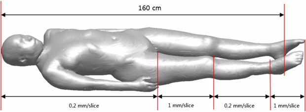
Figure 1. External form of female cadaver with indicated sectioning interval information.
Download figure:
Standard image High-resolution image2.2. Organ segmentation
The segmentation work was carefully performed in collaboration with professional anatomists. First, the slice images were taken at 2 mm intervals, and 825 images out of the total 5901 were selected for phantom construction. These images were then imported to Photoshop®CS4 (Adobe Systems, Inc., San Jose, CA) software for segmentation. Most of the organs were segmented manually with a screen digitizer (CINTIQ 15X, WACOM Co., Ltd, Japan); organs distributed throughout the body and clearly distinguishable, such as certain muscles and the blood, were segmented automatically using Photoshop®CS4's Color Range function. The skeletal system was also segmented automatically by CT imaging. Artifact-induced erratic structure segmentations were manually corrected. Figure 2 shows the example of a segmented image and a corresponding original color image.
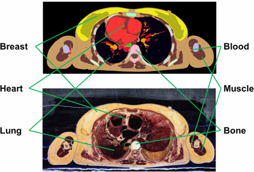
Figure 2. Example of segmented image with corresponding original color image.
Download figure:
Standard image High-resolution imageIn the present study, a total of 40 organs, including the 27 specified in ICRP Publication 103 (ICRP 2007), were segmented for effective-dose calculation. After segmentation, for memory-conservation and computation-speed purposes, the resolution of the images was reduced from 0.1 × 0.1 to 2 × 2 mm2. The resulting 2 × 2 × 2 mm3 voxel resolution provided sufficient accuracy for organ-dose calculation (Ai-dong et al 2006).
2.3. Adjustment of height and skeletal mass
The height and skeletal mass were adjusted by changing the voxel resolution. Specifically, the height of the unadjusted model (160 cm) was adjusted to the height of the reference Korean female (161 cm) by increasing the voxel size from 2 mm to 2.0747 mm in the longitudinal direction. With respect to the skeletal mass, as there is no available information in the reference Korean data, Clays et al (ICRP 1994)'s equation relating the total body weight to the total skeletal mass,

TBW being the total body weight in kg and SW the skeletal mass expressed as a percentage of TBW, were used. This equation yielded a total skeletal mass (including red bone marrow (RBM)) of 7.2 kg. Subsequently, this value was adjusted by changing the voxel size in the transverse direction from 2 × 2 to 2.0351 × 2.0351 mm2.
2.4. Weight adjustment for individual organs and whole body
After determining the voxel resolution, the masses of the individual organs and tissues were also adjusted to the reference Korean data. In the development of the HDRK-Man, the organs and tissues had been adjusted on 2D slice images using Photoshop 7.0TM's Inner Grow and Outer Grow functions. Due to the fact that the boundary voxels were only changed in the transversal direction, some organs (e.g. the spleen) were significantly deformed in appearance. In order to avoid this problem, in the present study, a 3D volume adjustment program was developed for boundary voxel change in 3D space. Checking the smoothness of an organ surface around a boundary voxel, the program decides boundary voxels to be changed (as shown in figures 3 and 4) in order to make an organ shape as smooth as possible. Conflicts such as organ overlap could be detected by the program and resolved manually. The program not only obviated a great amount of repetitive work, but also enabled closer matching of organ and tissue masses to the reference Korean data.

Figure 3. Procedures for decreasing an organ volume in the 3D volume adjustment program.
Download figure:
Standard image High-resolution image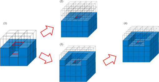
Figure 4. Procedures for increasing an organ volume in the 3D volume adjustment program.
Download figure:
Standard image High-resolution imageBecause some organs (RBM, oesophagus, bladder, ovaries, salivary glands, skin, adrenals, blood vessels, muscle, breast, colon, small intestine, uterus, thymus, eye lens, spinal cord, teeth, tongue, tonsils, trachea, bronchi, ureter, fallopian tubes, and vagina), had not been included in the reference Korean data, they were adjusted to the reference Asian data (IAEA 1998). Meanwhile, for organs heavier than those included in the reference Korean data, the volume was reduced by eliminating the surface voxels, which regions were replaced by adipose tissue. Conversely, for lighter organs, the volume was enlarged by changing boundary-region adipose tissue to organ material. All of the remaining volume in the phantom was defined as adipose tissue. After adjustment of the height and weight, skeletal mass, and organ and tissue masses, the phantom (54.8 kg) was slightly heavier than the reference Korean (54 kg); therefore, the weight was adjusted by eliminating adipose tissue from the surfaces of the legs. The average organ/tissue compositions in ICRU 46 (ICRU 1992) were used for all organs except the skeleton of which composition was used in ICRP 23 (ICRP 1975). The average organ/tissue densities in the reference Korean data (Park et al 2006), reference Asian data (IAEA 1998), and ICRU 46 (ICRU 1992) were used except for the lungs and skeleton. The lung density (0.392 g cm−3) was determined for the lung mass to be the reference lung value following the lung density determination method used in the ICRP reference phantoms (ICRP 2009). The skeletal density (1.347 g cm−3) was calculated based on whole skeleton density (1.3 g cm−3) reported in ICRP 70 (ICRP 1994), by excluding RBM (1.01 g cm−3).
2.5. Monte Carlo dose calculations
The developed HDRK-Woman was implemented in Monte Carlo particle transport simulation code MCNPX 2.7.0 (Pelowitz 2011) in order to enable calculation of organ doses for external photon exposures. The organ doses were calculated using the energy deposition tally based on track-length estimation (the F6 tally). Four photon-irradiation geometries were considered: anterior–posterior (AP), posterior–anterior (PA), right lateral (RLAT), and left lateral (LLAT). The calculated photon energies ranged between 0.015 and 10 MeV. The number of primary photons simulated was within the 108 (at 0.015 MeV)–107 (at 10 MeV) range considering statistical errors in the calculated values (i.e., to less than 10%, except for several results at 0.015 MeV for which the errors were unavoidably much larger). The computation time for each calculation case was 70–160 min on an AMD Opteron 6176 (2.3 GHz RAM, 64GB memory)-equipped server.
3. Results and discussion
3.1. HDRK-Woman
In the present study, a Korean adult female voxel phantom (HDRK-Woman; figure 5) was constructed using serially sectioned high-resolution color photographic images of a Korean female cadaver. The phantom is 161 cm in height and 54 kg in weight, which matches the figures in the reference Korean data. The voxel resolution is 2.0351 mm × 2.0351 mm × 2.0747 mm. The phantom is composed of 261 × 109 × 825 voxels ( = 23,470,425 voxels). The voxel phantom has 40 organs and tissues, including 27 organs for effective-dose calculations.
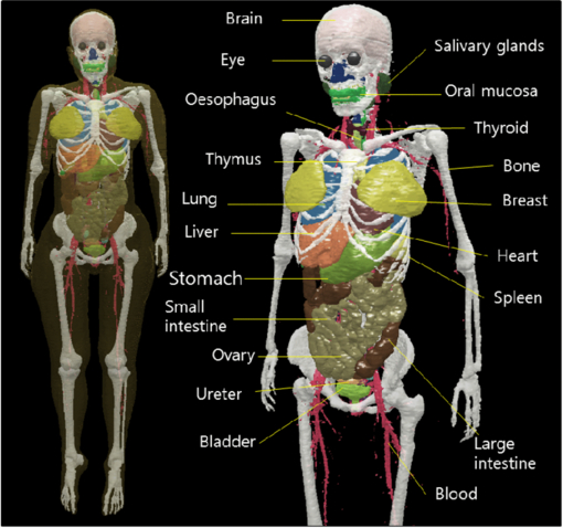
Figure 5. HDRK-Woman constructed in present study.
Download figure:
Standard image High-resolution imageTable 1 lists the phantom's organs and tissues alongside the reference Korean data. It is evident that the masses are closely matched to the reference Korean data, except for several organs, such as skin, blood, eye lens, tongue, trachea, and bronchi, due to the finite voxel resolution. The skin mass was 74.3% larger than that of the reference Korean data, even though the skin was defined as just a single voxel layer enveloping the phantom. The HDRK-Man and other phantoms, including the ICRP reference phantoms, exhibit the same problems. ICRP Publication 110 (ICRP 2009) indicates that several thin or small organs were not adjusted to match the reference values due to the finite voxel resolution.
Table 1. Numerical data for organs and tissue in the HDRK-Woman and the ICRP reference female phantom.
| Tissue | HDRK-Woman | ICRP female phantom | Reference | ICRP | |||
|---|---|---|---|---|---|---|---|
| Organ | weighting factor (wT) | Density (g cm−3) | Organ mass (g) | Density (g cm−3) | Organ mass (g) | Korean female (g) | Reference female (g) |
| Red bone marrow | 0.12 | 1.01 | 780.0 | – | – | 780a | 900 |
| (RBM) | |||||||
| Large intestine | 0.12 | 1.02 | 260.0 | 1.04 | 360.0 | 260a | 360 |
| Lungs | 0.12 | 0.392 | 693.0 | 0.384 | 950.0 | 693 | 950 |
| Stomach | 0.12 | 1.03 | 122.0 | 1.04 | 140.0 | 122 | 140 |
| Breast, adipose | 0.9 | 200.0 | 0.95 | 300.0 | 200a | 300 | |
| tissue | 0.12 | ||||||
| Breast, glandular | 1.02 | 300.0 | 1.02 | 200.0 | 300a | 200 | |
| tissue | |||||||
| Ovaries | 0.08 | 1.05 | 11.0 | 1.04 | 11.0 | 11a | 11 |
| Urinary bladder | 0.04 | 1.04 | 30.0 | 1.04 | 40.0 | 30a | 40 |
| Oesophagus | 0.04 | 1.02 | 30.0 | 1.03 | 35.0 | 30a | 35 |
| Liver | 0.04 | 1.06 | 1212.0 | 1.05 | 1400.0 | 1212 | 1400 |
| Thyroid | 0.04 | 1.05 | 12.0 | 1.04 | 17.0 | 12 | 17 |
| Bone | 0.01 | 1.35 | 6408.0 | 7760.1 | 6408 | 7760 | |
| Brain | 0.01 | 1.03 | 1403.0 | 1.05 | 1300.0 | 1403 | 1300 |
| Salivary glands | 0.01 | 1.03 | 62.0 | 1.03 | 70 | 62a | 70 |
| Skin | 0.01 | 1.08 | 3139.0 | 1.09 | 2721.5 | 1800a | 23 000 |
| Adrenals | 1 | 13.0 | 1.03 | 13.0 | 13a | 13 | |
| Extrathoracic (ET) | 1.03 | 41.0 | 1.03 | 18.61 | – | – | |
| region | |||||||
| Gall bladder | 1.03 | 17.0 | 1.03 | 10.2 | 17a | 8 | |
| Heart | 1.01 | 287.0 | 1.05 | 250.0 | 287 | 250 | |
| Kidneys | 1.05 | 290.0 | 1.05 | 275.0 | 290 | 275 | |
| Bloodb | 1.04 | 472.8 | 1.06 | 855.8 | 3800a | 4100 | |
| Muscle | 0.12 | 1.02 | 20 000.0 | 1.05 | 17 500.0 | 20 000a | 17 500 |
| Oral mucosa | 1.02 | 29.0 | 1.05 | 22.45 | – | – | |
| Pancreas | 1.05 | 58.0 | 1.05 | 120.0 | 58 | 120 | |
| Uterus/Cervix | 1.04 | 70.0 | 1.03 | 80.0 | 70a | 80 | |
| Small intestine | 1.02 | 450.0 | 1.04 | 600.0 | 450a | 600 | |
| Spleen | 1.06 | 153.0 | 1.04 | 130.0 | 153 | 130 | |
| Thymus | 1.01 | 29.0 | 1.03 | 20.0 | 29a | 20 | |
| Eye bulb | 1.03 | 19.0 | 1.05 | 15.0 | 19 | 15 | |
| Eye lens | 1.08 | 0.3 | 1.05 | 0.4 | 0.3a | 0.4 | |
| Spinal cord | 1.01 | 30.0 | 1.03 | 18.63 | 30a | – | |
| Teeth | 2.06 | 34.0 | 2.75 | 40.0 | 34a | 40 | |
| Tongue | 1.02 | 38.0 | 1.05 | 60.0 | 51a | 60 | |
| Tonsils | 1.03 | 3.0 | 1.03 | 3.0 | 3a | 3 | |
| Trachea | 1.06 | 12.0 | 1.03 | 7.99 | 68a | – | |
| Bronchi | 1.03 | 38.7 | 1.03 | 8.69 | 20a | – | |
| Ureter | 1.03 | 15.0 | 1.03 | 80.0 | 15a | 80 | |
| Fallopian tubes | 1.04 | 10.0 | – | – | 10a | – | |
| Vagina | 1.04 | 25.0 | – | – | 25a | – | |
| Adipose tissue | 0.9 | 15 376.7 | 0.95 | 23 596.4 | – | 22 500 | |
| Total body mass | 54 000.0 | 60 000.0 | 54 000 | 60 000 | |||
aThe reference Asian data (IAEA 1998) were used for the organs and tissues for which reference Korean data were not available. bBlood is used as a substitute to calculate lymph node doses due to the absence of lymph nodes in HDRK-Woman.
3.2. Comparison of organ doses (HDRK-Woman versus ICRP female reference phantom)
In the present study, organ doses or, more accurately, dose conversion coefficients (DCCs), defined as organ-averaged absorbed dose per photon fluence in the unit of pGy cm2, were calculated for photon exposures in the AP, PA, RLAT, and LLAT irradiation geometries. Then, the values obtained were compared with those of the ICRP reference female phantom. Note that the organ dose values of the first Korean female voxel phantom, KORWOMAN, are unavailable in literature for comparison. Figure 6 compares the DCCs for the values with relative errors (R) below 10%.

Download figure:
Standard image High-resolution image
Figure 6. Comparison of dose conversion coefficients (DCCs) between HDRK-Woman (filled) and ICRP female reference phantom (hole) for AP (squares), PA (circles), RLAT (triangles), and LLAT (diamonds).
Download figure:
Standard image High-resolution imageIt can be seen that the DCCs of the two phantoms are very close to each other if the photon energy is high (≥ 0.2 MeV). In fact, if only the high-energy photons (≥ 0.2 MeV) are taken into account, the average difference was 6.5% (standard deviation: 7.8%) for all organs and irradiation geometries. The maximum difference, 58.9%, was for the thyroid and the 0.2 MeV photon beam in the LLAT irradiation geometry.
If low-energy photons (≤ 0.08 MeV) also are considered, the difference is much larger. Indeed, for all of the organs and photon energies, the average DCC difference was 107.5% (standard deviation: 336.6%). The maximum difference, 2800%, was for the brain and the 0.015 MeV photon beam in the RLAT irradiation geometry.
Some discrepancies were found between the RBM and bone-surface (BS) DCCs. For the RBM, the HDRK-Woman phantom showed lower doses than the ICRP female reference phantom; however, for the HDRK-Woman phantom showed higher doses for the BS. This was owed to the HDRK-Woman and ICRP female reference phantoms' differing skeletal structures, which will be discussed in detail later.
The DCC discrepancies were mainly due to the organ-depth differences in the phantom. The importance of organ depth is acknowledged in ICRP (2009), which reported the ICRP reference phantoms' organ-depth distributions. In the present study accordingly, the HDRK-Woman phantom's organ-depth distributions were calculated for the AP, PA, RLAT and LLAT directions, and the distributions (determined by randomly sampling 107 points in the organs and calculating the distance of each from the phantom surface in a given direction) were compared with those in the ICRP female reference phantom. The results, plotted in figure 7, indicate significant organ-depth distribution differences between the phantoms. These differences explain most of the DCC inconsistencies between the HDRK-Woman and ICRP female reference phantoms; some of the DCC differences, however, had been influenced by other causes.

Download figure:
Standard image High-resolution image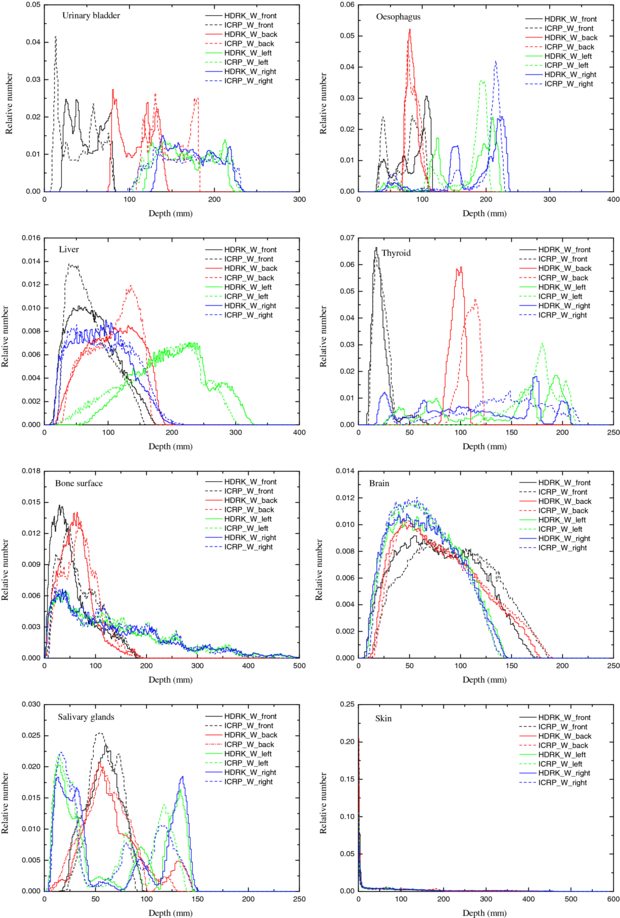
Figure 7. Comparison of organ-depth distributions for HDRK-Woman (straight line) and ICRP female reference phantom (dash line) in the skin below the body surface at front (black), back (red), left (green), and right (blue).
Download figure:
Standard image High-resolution imageIn the case of the RBM and BS for example, the DCC differences were mainly due to the different skeletal structures of the HDRK-Woman and ICRP female reference phantoms. The RBM of the HDRK-Woman was macroscopically defined by segmenting the red parts within the bones in the color images, similar to that of the HDRK-Man (Kim et al 2008), results show that the RBM is distributed deeply in the bones. On the other hand, it is not defined in the ICRP female reference phantom; instead, the spongiosa and medullary cavities surrounded by cortical bone, considered as one voxel layer, are used for RBM dose calculation purposes. The different skeletal structure of the both phantoms causes the calculated RBM doses for the HDRK-Woman to be lower than those for the ICRP female reference phantom regardless of irradiation geometry. The maximum difference, 55%, correlated with 0.03 MeV and the LLAT beam direction.
The BS, due to its small structure, was not able to be presented in the HDRK-Woman at the given voxel resolution, the same as in the HDRK-Man (Kim et al 2008). The BS doses for the HDRK-Woman were approximately estimated as the averaged dose for all of the bones (excluding the RBM), which are shielded mainly by muscle and adipose tissue, whereas those for the ICRP female reference phantom were calculated as the averaged dose to some parts of the spongiosa and medullary cavities, which are shielded not only by muscle and adipose tissue, but also by high-density cortical bone (1.929 g cm−3). Consequently, the BS doses for the HDRK-Woman were greater than those for the ICRP female reference phantom. These differences were much more severe for the BS than for the RBM: the maximum BS dose difference was about 1200% for 0.015 MeV and the AP beam direction. Note that Kramer et al (2007) already demonstrated that the BS dose approximation used in this study can overestimate BS dose values. Nevertheless, the overestimated BS dose values may insignificantly affect effective-dose values due to the low BS weighting factor at 0.01 (ICRP 2007).
With respect to the brain, the dose difference was mainly due to the density difference between the cranial bones: 1.35 g cm−3 for the HDRK-Woman and 1.505 g cm−3 for the ICRP female reference phantom. The brain doses for the HDRK-Woman were, therefore, greater than those for the ICRP female reference phantom for all beam directions. The maximum difference, as large as about 2800%, corresponded to 0.015 MeV and the RLAT beam direction.
To elucidate the correlation between the organ-depth distributions and the DCCs, the average values of the organ-depth distributions and the natural logarithms of the dose values calculated for 0.03 MeV photons were plotted in a scatter diagram (figure 8). The circles and triangles represent the HDRK-Woman and ICRP female reference phantoms, respectively. A negative linear association between the two variables is apparent for both phantoms. To evaluate the statistical significance of that association, the variables' correlation coefficient was calculated, and a statistical hypothesis test was performed under the significance level of 0.05 using t-statistics. As determined, the correlation coefficient was −0.795; the p-value was less than 0.001, much lower than the significance level. These results indicated that the negative linear association was statistically significant.
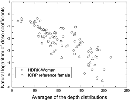
Figure 8. Scatter diagram of average of depth distributions and natural logarithm of dose coefficients (for 0.03 MeV photons) for HDRK-Woman (circles) and ICRP female reference phantom (triangles).
Download figure:
Standard image High-resolution image3.3. Comparison of effective doses (HDRK phantoms versus ICRP reference phantoms)
The effective doses calculated using the HDRK phantoms (HDRK-Man and HDRK-Woman) were compared with those determined with the ICRP reference phantoms reported in ICRP Publication 116 (ICRP 2010). Figure 9 plots the effective-dose conversion coefficients for the four irradiation geometries considered in the present study. It can be seen that the difference was almost negligible when the photon energy was equal to or greater than 0.08 MeV. The average difference was 4.9% (standard deviation: 4.5%); the maximum difference was 17.1%, which correlated with 0.6 MeV and the RLAT direction. When the low-energy photons (≤0.05 MeV) were also considered, the difference was larger: 18.6% (standard deviation: 21.6%); the maximum difference was 76% for the 0.015 MeV beam and LLAT direction.

Figure 9. Comparison of effective-dose conversion coefficients (ECCs) between HDRK phantoms and ICRP reference phantoms.
Download figure:
Standard image High-resolution image
4. Conclusions
In the present study, a Korean adult female voxel phantom, HDRK-Woman, was constructed using high-resolution color photographic images of a Korean adult female cadaver. The dimensions of the phantom, including the body height and weight, the skeletal mass, and the dimensions of the individual organs and tissues, were adjusted to the reference Korean data. The organ doses (more accurately: dose conversion coefficients or DCCs) for external photon exposures and organ-depth distributions were calculated and compared with those obtained with the ICRP female reference phantom. Additionally, the effective-dose conversion coefficients (ECCs) were calculated using both the HDRK-Man and HDRK-Woman, and the determined values were compared with those of the ICRP male and female reference phantoms. The results showed that the ECCs are indeed significantly different if the photon energy is very low (≤0.05 MeV), meaning that differences of anatomical structure should be considered at least for low-energy photons. For high-energy photons, which are more frequently employed, the ECC differences (average: 4.9%) were very small. These, considering the dose calculation uncertainties in the radiation protection realm, generally can be ignored. The HDRK-Man and HDRK-Woman can be used for dose calculation for Korean workers in the future. Note that the HDRK phantoms have some limitations including: (1) significant overestimation of the bone surface (endosteum) doses and (2) absence of lymph nodes of which doses are inevitably substituted by blood doses. In a future study, we will update both the HDRK-Man and HDRK-Woman in order to address these limitations. Also note that the HDRK phantoms, because their dimensions are adjusted to the reference Korean, were only designed for radiological protection purposes and cannot be used in medical applications for which patient-specific phantoms are required.
Acknowledgments
This research was supported by the General Research Program, the National Nuclear R&D Program and Global PhD Fellowship Program through the National Research Foundation of Korea (NRF) funded by the Ministry of Science, ICT and Future Planning (nos NRF-2011-0025496, NRF-2012-K001146, NRF-2012M2A8A5026057, NRF-2011-0007318 and 2011-0030970).