Abstract
We provide direct evidence of plasma-induced pore formation in a cell membrane model system. We irradiated plasma on the basis of the dielectric barrier discharge onto a supported lipid bilayer (SLB). Observation with a fluorescence microscope and atomic force microscope revealed the formation of pores on the order of 10 nm–1 µm in size. Capturing these micropores in a fluid lipid membrane is a significant advantage of the SLB system, and quantitative analysis of the pores was performed. Stimulation with equilibrium chemicals (HNO3 and H2O2) indicated that other transient active species play critical roles during the poration in the SLB.
Export citation and abstract BibTeX RIS

Content from this work may be used under the terms of the Creative Commons Attribution 4.0 license. Any further distribution of this work must maintain attribution to the author(s) and the title of the work, journal citation and DOI.
This article was made open access on 16 September 2015
Atmospheric plasma is applied as a novel and valuable tool in the medical and biological fields.1–3) Recent studies have demonstrated that the plasma irradiation of cells or organs is applicable multifariously, including sterilization,4–8) selective killing of tumor cells,9) cellular regulation,10,11) and wound healing.12) In spite of the wide variety of medical and biological applications of plasma, many aspects remain unclear, including what parts of plasma affect cells and tissues and how they do so. Atmospheric plasma generates various active species called reactive oxygen and nitrogen species (ROSs and RNSs) including acidic and radical species,6–8) which transfer to cells through the medium around the cells. The critical effect of these active species on cells may be a direct effect on cell surfaces or an indirect one through physiological cascades inside cells and/or genetic damages. Biological cell membranes, also called plasma membranes, act as barriers to maintain the integrity of cells by separating the inside from the outside. Chemically active species produced by plasma irradiation first access the cell membranes. Some recent studies showed that cell membranes act as a barrier against plasma irradiation for genomic damage or oxidative protein modification,13,14) while other studies showed that plasma irradiation causes the oxidation of the intracellular organelles because of the permeation of ROS through the cell wall and cell membrane, without a major deformation of the membranes.6,15) How the plasma-induced active species affect and/or permeate cell membranes is unclear. Physical damage to a cell membrane, such as poration, is effective for the transportation of solutes including plasma-induced active species into cells, but such a non-selective leakage itself is highly toxic for cells, similar to pore-forming proteins.16) Permeation via dissolution into the hydrophobic core of a cell membrane is inefficient for the water-soluble active species and possibly prevented by drug efflux pumps.17) The lipid peroxidation induced by physiological ROSs is also related to cell damage and various pathological states.18–20) A fundamental understanding of the effects of plasma on biomolecules and their assembled structures is demanded for the further progress and establishment of safety in the medical and biological application of plasma.
Artificial lipid bilayers are useful biomembrane model systems for investigating the fundamental interaction between cell membranes and medical and biological agents.21–25) The artificial planar lipid bilayers formed at solid–liquid interfaces are called supported lipid bilayers (SLBs), and the spherical lipid bilayers dispersed in aqueous solutions are called lipid vesicles. SLBs have the advantages that fluid and fragile lipid membranes exist stably owing to the support of the solid substrate and that high-resolution surface scientific techniques such as atomic force microscopy (AFM) are adopted. Recently, Svarnas et al. reported the plasma irradiation of a suspension of multi-lamellar vesicles (MLVs),26) which is the only example of plasma irradiation of an artificial lipid bilayer, to our knowledge. The plasma irradiation induces the fusion of MLVs, but the leakage of the encapsulated solution in MLVs is limited.26)
In this study, we investigated the effect of the atmospheric plasma on the basis of the dielectric barrier discharge (DBD) on an artificial cell membrane using the SLB system. We visualized that the irradiation of the DBD-plasma to an SLB of phospholipid-generated pores with a diameter on the order of 10 nm–1 µm. The pore formation proceeded without a change in pH, a comparison with the effects of the chemicals in the equilibrium condition indicated the critical roles of the plasma-induced transient active species.
A schematic of the DBD-plasma irradiator developed for this study is shown in Fig. 1(a). A quartz plate was fixed on each of the upper and lower electrodes, which were used to apply an AC voltage. A sinkhole was fabricated on the lower quartz plate and used as a dish for the SLB formation, DBD-plasma irradiation, and observation on the SiO2/Si substrate. The distance between the upper and lower quartz plates was 1.5 mm. The thickness of the buffer solution above the SiO2/Si substrate was ∼0.47 mm, and the solution was carefully treated so as not to expose the SiO2/Si surface to the air, because the SLB stably exists only in an aqueous solution. We settled the DBD-plasma irradiator in a glove box and purged its inside with Ar for 5 min before applying an AC voltage of 15 kV and 15 kHz between the electrodes to generate the DBD-plasma. The plasma was irradiated to the SLB on the SiO2/Si substrate for 30–150 s through the buffer solution. The increase in the temperature due to the plasma irradiation was suppressed by the circulated coolant in the electrodes. The DBD in the Ar gas ambient, which probably included a slight amount of residual air, contained rapidly moving bright filamentary discharges [Fig. 1(b)]. The details of the instrument are described elsewhere.27,28)

Fig. 1. (a) Schematic of the instrument and sample setup of the DBD-plasma irradiation of an artificial lipid bilayer membrane on a SiO2/Si substrate through a buffer solution. (b) Photograph of the DBD-plasma irradiation.
Download figure:
Standard image High-resolution imageWe used dioleoylphosphatidylcholine (DOPC) because phosphatidylcholine is the representative phospholipid most abundantly existing in cell membranes. A vacuum-dried film of DOPC mixed with a fluorescence-labeled lipid [dioleoylphosphatidylethanolamine-N-(lissamine rhodamine B sulfonyl) (Rb-DOPE)] at the molar ratio of  was prepared from their chloroform solutions and suspended in a buffer solution [100 mM KCl, 25 mM HEPES/NaOH (pH 7.4)] at the lipid concentration of 0.40 mM to prepare MLVs Lipids and other chemicals were purchased from Avanti Polar Lipids and Wako Pure Chemical Industries, respectively. The MLVs were transformed into unilamellar vesicles with an average diameter of 120 nm through freeze-and-thaw cycles and an extrusion process using a 100-nm-pore polycarbonate filter.29–31) The DOPC-SLB containing Rb-DOPE was prepared on thermally oxidized SiO2/Si substrates using the vesicle fusion method.32) The SLBs before and after the plasma irradiation were observed using an epi-fluorescence microscope (epi-FM; Olympus BX51WI) and an AFM (Agilent PicoPlus 5500, formerly Molecular Imaging). We kept the SLB samples under the buffer solution during the entire preparation, plasma irradiation, and observations. Further details of the sample preparation and observations are described elsewhere.29–31) We used Image J software (NIH, http://imagej.nih.gov/ij/) for the analysis of epi-FM images. The area fraction and density of objects were evaluated as the average of at least four images for each experimental condition. The area fraction in the SLB before the plasma irradiation, which contained several defects in some parts, was also measured and subtracted from that after the plasma irradiation.
The epi-FM images of the DOCP-SLB on the SiO2/Si substrate before and after the DBD-plasma irradiation are shown in Fig. 2. The majority of the substrate was covered by a defect-free SLB [Fig. 2(a)]. We confirmed using fluorescence recovery after the photobleaching method that the DOPC-SLB was fluid and continuous in the field of view.30,31) Figure 2(b) shows the DOPC-SLB after the DBD-plasma irradiation for 150 s; dark regions appeared in the DOPC-SLB [Fig. 2(b)]. We attribute the dark regions to pores generated in the lipid bilayer membrane by the DBD-plasma irradiation. The other possible cause of the dark regions is lipid domains, excluding the dye-labeled lipid in the case of a multi-component system,33) but we omit this possibility in the single-component SLB system in the present study. Regarding the plasma irradiation conditions for Fig. 2(b), the density of the defect was 9.3 × 10⁻³ µm−2, which corresponded to ∼1 defect per 10 × 10 µm2 area, and the area fraction of the pores (θpore) was 3.1%. We confirmed that the pores were not formed by the temperature increase up to 70 °C or the application of the AC voltage without discharge.

Download figure:
Standard image High-resolution image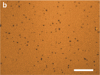
Fig. 2. Fluorescence images of the DOPC-SLB (a) before and (b) after the plasma irradiation for 150 s. Scale bars: 20 µm.
Download figure:
Standard image High-resolution imageThe dependence of θpore on the time of the plasma irradiation is shown in Fig. 3. The generation of pores was not significant until the irradiation time of 30 s, but θpore increased with time for irradiation times longer than 60 s. Note that the values of θpore in Fig. 3 were obtained from optical microscope images; thus, the pores smaller than the diffraction limit were not detected. It is possible that pores existed that were too small to be detected using a conventional fluorescence microscope.

Fig. 3. Dependence of the plasma-induced θpore on the irradiation time evaluated using fluorescence images.
Download figure:
Standard image High-resolution imageTherefore, we investigated the surface morphology of the DOPC-SLB on the submicron scale using AFM (Fig. 4). The DOPC-SLB surface before the plasma irradiation was flat, with a roughness of 0.23 nm (rms) [Fig. 4(a)], as in a previous study.31) After the plasma irradiation, small pits appeared on the SLB surface [Fig. 4(b)]. Figure 4(c) shows the phase-shift image obtained simultaneously with Fig. 4(b) to facilitate recognizing the position of the pits. We assign these plasma-induced pits to the initial state of the micrometer pore formation observed using the epi-FM in Fig. 2(b). We recognized 41 pits in 1 µm2 of Figs. 4(b) and 4(c), and their lateral and vertical average sizes were 16.4 ± 4.4 nm in diameter and 0.81 ± 0.19 nm in depth, respectively. If we assume a curvature radius of 10 nm for the tip of the cantilever, the depth of the pits was possibly underestimated because the cantilever tip did not reach the bottom of the pits. The actual depth may be larger than 0.81 nm, which is why the pit depth was smaller than the thickness of the DOPC-SLB (∼4 nm).32) The diameter of the pits is hardly affected by the tip shape. It should be noticed that the tip-shape effect for depressions is different from that for protrusions, in which height is measured correctly but diameter is overestimated.34)

Fig. 4. AFM topographies (1 × 1 µm2) of the DOPC-SLB (a) before and (b) after the plasma irradiation for 120 s. Cross-section profile of the white line is also shown in (b). Blue arrows indicate the same plasma-induced pit. (c) Phase-shift image simultaneously obtained with (b).
Download figure:
Standard image High-resolution imageThe epi-FM (Fig. 2) and AFM (Fig. 4) images provided direct evidence of the pore formation due to the DBD-plasma irradiation. We emphasize that it is owing to the SLB system that the small pores on the order of 10 nm [Figs. 4(b) and 4(c)]–1 µm [Fig. 2(b)] in size were captured and visualized successfully. Pores in cell membranes and free-standing membranes such as giant unilamellar vesicles recover to a continuous membrane35) because the edge of lipid bilayers is energetically unfavorable.36,37) On the other hand, it is well known that pores and edges exist stably in SLB systems.30,31) We attribute the formation of nanopores by the DBD-plasma irradiation [Fig. 4(b)] to the initial plasma effect on the biomembranes. A quantitative analysis of the relation between the plasma irradiation and pore formation was achieved using the SLB system, as shown in Fig. 3. The pore formation itself causes damage to cells because of the non-selective permeation of solutes into and out of cells, as well as the easier transfer of ROS and RNS into the cytosol and nucleus. Additionally, the oxidation of lipids affects the lipid domains38) and thus the functions of membrane proteins. We notice that the plasma-induced nanopores were still metastable, even in the SLB system, and were scarcely found 1–2 h after the plasma irradiation during the sequential AFM experiments. Statistical and quantitative analysis of the initial rate of the pore formation by using AFM is remained for future studies.
We investigated the effects of chemical stimulations to the DOPC-SLB and compared them with the results of the plasma irradiation. Nitrate ion (NO3−) and hydrogen peroxide (H2O2) are representative byproducts generated by plasma-induced RNSs and ROSs.6–8) Figures 5(a) and 5(b) show the DOPC-SLB after the addition of nitric acid (HNO3) at final concentrations of 37 and 183 mM, respectively. Dark defect regions appeared after the addition of nitric acid at the concentration of 37 mM [Fig. 5(a)], but further addition of HNO3 did not increase the defects [Fig. 5(b)]. The estimated pH values in Figs. 5(a) and 5(b) are 1.4 and 0.7, respectively. We attribute the HNO3-induced defect formation to the effect of the low pH near 1 as the previous report,39) in which the defect formation is explained as the budding and rupture of the bilayer membrane due to the electrostatic interaction. The HNO3-induced defect formation was independent of the HNO3 concentration and thus different from the tendency of the DBD-plasma-induced defect generation, as shown in Fig. 3. Similar defects were also generated by sulfuric acid (H2SO4) [Fig. 5(c)]. It was reported that the concentration of the plasma-induced NO3− or NO2− is on the order of 10 nM.6) Even the excess amount of HNO3 in Fig. 5(a) only caused the pH-induced effect to SLB and did not exhibit HNO3-specific effects on the SLB. Therefore, we conclude that the plasma-induced poration in the DOPC-SLB shown in Figs. 2 and 4 was not because of a specific effect of NO3− on lipid molecules. Using pH paper, we confirmed that the DBD irradiation of the buffer solution in the same conditions as that for SLB (700 µL of the buffer solution, for 150 s) did not change the pH. Figure 5(d) shows the epi-FM image of the DOPC-SLB after the addition of H2O2. The addition of H2O2 at the maximum concentration of 248 mM caused little morphological change, and we did not observe the generation of any defects [Fig. 5(d)].

Fig. 5. Fluorescence images of the DOPC-SLB after the addition of chemical agents. (a) HNO3, 37 mM; (b) HNO3, 183 mM [the same position as (a)]; (c) H2SO4, 153 mM; and (d) H2O2, 248 mM. Scale bars: 20 µm.
Download figure:
Standard image High-resolution image
The results shown in Fig. 5 indicate that the active species for the pore formation in the DOPC-SLB were not the chemical species in an equilibrium state such as NO3− or H2O2 but other transient ROSs or RNSs.8) Recent studies indicated that plasma sterilization proceeds at pH lower than 4.7 and that the hydroperoxy radicals (HOO ), which exist predominantly at pH <4.8, play a key role in the sterilization.6,7) We propose that they are effective for the proteins or genes in cytosols and nuclei but not for the poration in cell membranes, on the basis of our present results indicating that the defects in the DOPC-SLB were formed without pH change. The area fraction of the defects generated by the DBD-plasma was ∼3% at maximum after the plasma irradiation for 150 s, even in the SLB system (Fig. 3). If we consider the number density, the majority of the plasma-induced defects were nanopores of ∼10 nm diameter, as observed using AFM (Fig. 4). These pores are sufficiently large for the non-selective leakage of solutes into and out of cells, considering the sizes of pore-forming proteins,16) but their lifetime is short in a self-healing membrane system, as discussed above. This is one of the reasons why the plasma-induced damage is limited in cell membranes compared with in the proteins and genes of cells.6,13,15) In the case of MLV, the poration is limited only at the outermost few layers and does not reach the inner shells.26) Further studies are needed to identify the critical species of the pore formation in lipid bilayer membranes among the numerous candidates for plasma-induced ROSs and RNSs,8) but the SLB system is valuable for the investigation of the effects of plasma on biomembranes.
In conclusion, we provided fundamental information about the effect of atmospheric plasma on a lipid bilayer membrane. DBD-plasma irradiation of a DOPC-SLB generated 10 nm–1 µm micropores without a change in pH. We propose that SLBs are effective for obtaining information about the physical and chemical modification of cell membranes induced by plasma.
Acknowledgments
We express gratitude to Ryuma Yamashita, Toyohashi University of Technology, for the contribution and support of the experiments. This work was supported by KAKENHI Grant Numbers 23685003, 24360108, 25630110, and 24110708, "Program to Foster Young Researchers in Cutting-Edge Interdisciplinary Research" from JST, the EIIRIS Project from Toyohashi University of Technology.