


Abstract
Recent advances in the manipulation of molecules now allow us to also probe nanoporous silified biomaterials. We demonstrate the quantum coherent propagation of phthalocyanine through the skeleton of the alga Amphipleura pellucida. A micro-focused laser source prepares a molecular beam which is sufficiently delocalized to be coherently transmitted through the alga's frustule—in spite of the substantial dispersive interaction between each molecule and the nanomembrane.
Export citation and abstract BibTeX RIS

Content from this work may be used under the terms of the Creative Commons Attribution 3.0 licence. Any further distribution of this work must maintain attribution to the author(s) and the title of the work, journal citation and DOI.
GENERAL SCIENTIFIC SUMMARY Introduction and background. Physics associates all quantum matter with wave properties. Even almost 100 years after the introduction of this concept by Louis de Broglie, this phenomenon still simulates discussions among scientists and philosophers when it comes to matters of high complexity since it questions our common-sense understanding of reality or space–time. We usually don't perceive it since the de Broglie wavelength shrinks with the object's mass and velocity. Quantum interferometry therefore usually requires costly nanotechnology.
Main results. Here, we show for the first time that quantum coherence is sufficiently robust and biological grown nanostructures are sufficiently small and regular to allow the quantum coherent transport of individual dye molecules through the open pores of the silified skeleton of a marine alga suspended in vacuum. Amphipleura pellucida is part of the marine phytoplankton which you can collect on the beach. With a wall thickness of 90 nm and a surprisingly regular pore distance of about 200 nm it allows us to measure de Broglie wavelengths as small as a few billionths of a millimeter by quantum diffracting molecules at the biologically grown nanomask.
Wider implications. The Vienna experiment prepares macromolecular quantum interference for the first time with means that are in reach for almost every undergraduate physics lab. Moreover, Amphipleura pellucida is by no means a special case. Several tens of thousands of similar algae are waiting to be collected at the beach. Future studies of quantum diffraction are also expected to reveal information on the inner pore structure via phase tomography—complementary to the topography that is readily obtained in electron microscopy.
 Figure. The quantum coherent propagation of individual PcH2 molecules through the alga's pores is revealed by the appearance of matter-wave interference fringes on the detection screen. The molecules are launched by laser microevaporation and detected in fluorescence imaging.
1. Introduction
Throughout the last few decades, matter–wave physics has probed fundamental quantum physics [1–4] and has led to interesting applications in precision measurements, quantum sensing and quantum information.
Recently, it has become possible to extend the range of coherent manipulation schemes to the de Broglie interference of complex molecules [5–8], including quantum-enhanced molecule metrology [9, 10] and coherent nanoparticle nanolithography [11].
For massive matter, one of the conceptually most elementary gedanken-experiments of quantum physics—the diffraction at a double slit or grating—usually implies rather sophisticated micro- or nanotechnologies to realize diffraction elements that are adapted to very short de Broglie wavelengths [12–17].
In this work, we emphasize that molecular coherence experiments can also be done with simple structures made by nature, and that molecular diffraction is interesting and well adapted for the imaging of nanoporous structures. Particles of mass m and velocity v are associated with a short de Broglie wavelength λdB = h/mv. They achieve high sensitivity to external potentials and internal phase changes, at rather low kinetic energies. Molecules are also interesting nanoprobes, since we are free to choose them according to their internal characteristics—with different electric, magnetic or optical properties. The high fluorescence yield of PcH2 makes it a particularly appealing candidate for particle detection at the single-molecule level.
We exploit here a coherent molecular beam of phthalocyanine to obtain a one-dimensional (1D) Fourier projection of the silified cell wall (frustule) of the diatom Amphipleura pellucida. These unicellular algae, which can be found in large abundance in freshwater phytoplankton, have attracted great interest in the nanotechnology [18–20] and optics community [21, 22].
Van der Waals forces are thought to have a high impact on the performance of algae as nanoporous filters [23]. In our experiments, we can access these forces, as they directly affect the observed interference patterns.
2. Experimental setup
The experiment is sketched in figure 1(A). It consists of three principal components: a coherent molecular beam source, the frustule of a diatom—suspended in vacuum with its pores oriented toward the molecular beam—and a laser-induced fluorescence microscope with single-molecule sensitivity.
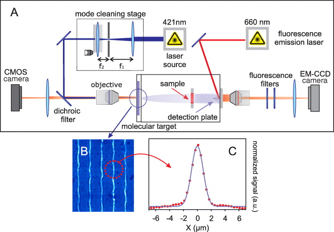
Figure 1. (A) Experimental setup: after launch in a microfocus laser evaporation source (421 nm, 100 mW diode laser), the molecular beam coherently illuminates the frustule of the diatom Amphipleura pellucida. The interference pattern is collected on a quartz plate and imaged using single-molecule fluorescence microscopy, excited by a red diode laser (661 nm, 12 W cm−2) [24]. (B) Light microscopy picture of some desorbed lines from the molecular target. (C) The cross-cut shape of the lines is approximately Gaussian with a waist of 1.6 μm.
Download figure:
Standard image High-resolution imageIn order to observe an interference pattern, we first need to prepare the transverse coherence of the molecular de Broglie wave by reducing the source size to a level, such that quantum indeterminism ensures the required coherent momentum spread and the position delocalization across several alga pores. We have implemented this idea using a micro-focus laser evaporation source. A 421 nm laser beam (70 mW) is spatially filtered with a 15 μm pinhole and tightly focused by a 50 × objective onto a vacuum window whose inner surface is coated with many layers of PcH2 (514 amu). The molecules are thus heated and evaporated into high vacuum (10−8 mbar). The window is mounted onto a motorized translation stage and it is continuously scanned at 1 mm s−1 to expose new material to the laser beam. The evaporation process is monitored by imaging the emitted source fluorescence onto a CMOS camera. Figure 1(B) shows a picture of the molecular target: five lines desorbed from the substrate are depicted, each with a Gaussian waist of roughly 1.6 μm (figure 1(C)). The high localization on the glass and Heisenberg's uncertainty relation then guarantee the coherent illumination of the diatom sample 1.57 m downstream.
The micro-laser source has many benefits: it can be operated in a pulsed mode on a short time scale to protect the thermo-labile molecules. It can serve as a multi-species source, with different molecules coated onto different regions of the same substrate. The evaporation region can also be arbitrarily shaped (point, slit, circle, square) to the needs of the experiment.
Figure 2 shows scanning electron microscope (SEM) images of the alga. Its boat-like shape is structured by a natural array of nanopores with an average distance of 205 nm and a typical width of 122 nm along the x-axis. According to electron energy loss spectroscopy measurements, the cell wall is roughly 90 nm thick and suspended between rod-like support structures of 110 nm thickness.
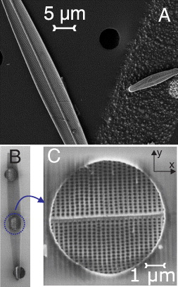
Figure 2. SEM pictures of Amphipleura pellucida. (A) The diatom has a boat-like shape. (B) The frustule is masked by a collimation hole of 5 μm diameter. (C) The diatom frustule fully covers the selected hole. The nanoporous arrays were aligned to within 5 mrad with respect to gravity.
Download figure:
Standard image High-resolution imageAn aqueous solution of diatoms was gently dropped onto a holey carbon grid. The drying process results in a random distribution of diatoms that are electrostatically bonded to the carbon mesh. We have selected a diatom that fully covered one of the 5 μm holes in the membrane and we blocked all remaining holes with a 50 μm thick mylar foil. Selecting a circle of 5 μm diameter is necessary in order to reduce the divergence of the molecular beam to less than the expected diffraction angle.
Quantum diffraction of the coherent molecular beam at the alga's nanoporous structure leads to the formation of an interference pattern, i.e. a molecular density pattern that is deposited on a quartz window 0.57 m further downstream at the vacuum–air interface. It is imaged in situ and with single-molecule sensitivity using fluorescence microscopy. The laser evaporation source generates a molecular velocity distribution that can be fitted with a Maxwell–Boltzmann curve at a temperature of ∼ 675 K. Since the molecules fall freely in the gravitational field, fast molecules end up at the top and slow molecules at the bottom of the detection screen (figure 3(A)). We can thus assign a molecular velocity band to a given height on the detector.

Figure 3. (A) Diffraction pattern of PcH2 molecules after their coherent propagation through the frustule of Amphipleura pellucida. The vector of gravity is oriented vertically. (B) Predicted 2D interference pattern based on the binarized version of figure 2(C), not including gravity or any additional phase shifts. (C) Vertically integrated trace of the experimental 2D diffraction image (A) in the mentioned velocity range (red dots). The higher-order, high-contrast interference fringes document the high molecular coherence as well as the presence of interactions between the phthalocyanines and the alga's frustule. While the higher orders are missing in the simple simulation (B), they can be qualitatively reproduced (panel C, green line) by assuming an effective radial reduction of the pore openings by 83 nm in the 2D simulation of panel (D). The simulation represented by the black line in (C) starts from the measured transmission mask (figure 2(C)) and includes a basic 1D horizontal van der Waals dephasing [25] with C3 = 66 nm3 meV.
Download figure:
Standard image High-resolution image
3. Discussion
Using the SEM image of the alga's frustule (figure 2), we can predict the two-dimensional (2D) diffraction pattern in the absence of gravity (figure 3(B)). To do so, we first binarize the porous structure and calculate the interference pattern by solving the Kirchhoff–Fresnel integral, including the finite source size and the velocity spread of the molecular beam. Although the nanoporous array was aligned to within 5 mrad with respect to the axis of gravity, we expect the vertical diffraction peaks to smear out because of the free-fall spread, while the horizontal diffraction fringes will be fully preserved. The expected result is the blue solid line in figure 3(C). We compare this with the experimental result by integrating the 2D data of figure 3(A) vertically over the same velocity band of v = 142–168 m s−1. This interference line is represented by the red dots of figure 3(C). We can draw a number of conclusions from these curves.
Firstly, the high interference fringe visibility proves the coherent transport of material through the silified microbiological specimen. It is fully consistent with assuming each molecule to be delocalized across several of the alga's pores.
Secondly, by evaluating the distance between the zeroth and the first diffraction peak, we are able to reconstruct the expected pore periodicity of 205 nm with an accuracy of ∼ 5%.
Thirdly, the comparison between the naive Kirchhoff–Fresnel solution and the experiment shows that we observe more and more intense diffraction peaks than the simulation would suggest. It has been known from earlier experiments [24–26] that the van der Waals interaction between polarizable particles and a nearby solid surface induces a significant phase shift in the diffraction process. The most prominent overall effect of this interaction is to virtually reduce the open pore size. This is corroborated by figure 3(D), which shows how the higher diffraction orders are populated when we reduce the pore size in our simulation. The vertically integrated trace of that is shown as the green solid line in figure 3(C).
We may also model the effect by integrating a van der Waals phase shift with a fixed C3-constant, where we only take into account the horizontal particle–wall distance in the cylindrical pores. The result of that is shown as the black solid line, which also fits the experimental data reasonably. Molecular diffraction can yield information of the internal material properties of a mechanical masks. A fully quantitative model will, however, have to include various details such as entrance and departure effects, pore irregularities, the full electronic spectrum of the molecules and others.
4. Conclusion
Our proof-of-concept experiment demonstrates high-contrast coherent diffraction of molecules at a biological nanostructure. Future studies shall also explore 2D-holography of different types of diatoms in a vertical setup. This shall permit one to not only reveal the structural differences between frustules of different alga specimens but also to distinguish different internal properties, which affect the molecular phase shifts. More than 100 000 different types of diatoms are known to exist [27] and also offer a playground for a number of pedagogical experiments in matter–wave physics.
While natural diatoms have a silica frustule, it has been shown that it is also possible to systematically exchange the diatom's base material. In future experiments, it could be intriguing to explore arbitrary but topologically equal shapes with different atomic compositions [28] (Si, Ge, Mg) to shed more light on van der Waals interactions in natural geometries.
Acknowledgments
We acknowledge the financial support by the FWF funds under contract Z149-N16 (Wittgenstein) and by the European Commission under contract FP7 304886 (Nanoquestfit). We acknowledge the discussions with I Gebeshuber on diatoms and S Scheel, J Fiedler, S Nimmrichter on modeling. We thank S Puchegger for his support in SEM imaging and J Bernardi for his support in STEM measurements at USTEM, Vienna.