
Abstract
Exhibiting rich magnetic behaviour and potentially multiferroic properties, the dugganites, a Te6+ containing subgroup of the langasite series, are an attractive family of compounds for future study. It was recently shown that Pb-bearing members of the dugganite series undergo distortions away from the P321 symmetry that is characteristic of the langasites. Here, we detail the consequences these distortions have on the magnetism exhibited by Pb3TeCo3V2O14 and Pb3TeCo3P2O14, solving the magnetic structures of both compounds with respect to a new supercell. Using neutron scattering and magnetic susceptibility measurements, we show that small applied magnetic fields can seriously perturb the delicate magnetic states in both of these systems. This is further demonstrated by presenting how doping P5+ onto the nonmagnetic V5+ site completely changes the magnetic structure from either of the end series members. Finally, it is shown using inelastic neutron scattering and magnetic susceptibility measurements that Pb3TeCo3V2O14 can be characterized using a model for isosceles trimers, which do not exist in the previously reported P321 subcell.
Export citation and abstract BibTeX RIS
1. Introduction
Composed of well over 100 different species, the langasites are an extremely diverse class of compounds that have attracted the attention of materials scientists from many disciplines. This is mainly due to their general ease of crystal growth [1–4], structural diversity [5], and wide range of behaviours including piezoelectric [6], optical [7], dielectric [8], and multiferroic properties [9, 10]. In addition, the frustrated nature of the magnetic sublattice presents an opportunity to study interesting magnetic phases including doubly-chiral magnetic structures [11–13], or the exploration of spin-liquid dynamics at low temperatures [14, 15]. The typical langasite has the Ca3Ga4Ge2O14 structure: a trigonal unit cell with noncentrosymmetric P321 space group, which is largely responsible for many of the previously listed behaviours. However, occasionally langasites may deviate from this symmetry either through spontaneous or pressure-induced phase transitions [5].
Recently, a subclass of the langasites, called the dugganites (named after the mineral dugganite Pb3TeZn3As2O14 [16–18]), has been characterized. These materials all contain Te6+ at the 1a octahedral site, which has important implications for the magnetism of the dugganites. Unlike other ions which commonly occupy the 1a site such as Ga5+ and Nb5+, Te6+ is only known to exist in octahedral coordination [19]. This forces other ions like Co2+, which often prefers the octahedral site, solely into the 3f tetrahedral site. Atoms occupying the 3f site are arranged in isolated trimeric units which stack along the c axis [10]. Pb3TeCo3V2O14 was recently introduced as a potential multiferroic candidate displaying not one, but two exotic magnetic transitions [20]. Even more recent was the discovery of a monoclinic superstructure which occurs on nearly all Pb-containing dugganites characterized to date, except maybe for the natural mineral [21]. Here, we report how this supercell influences the magnetism and spin dynamics in Pb3TeCo3V2O14, Pb3TeCo3P2O14, and doped series members Pb3TeCo3V2−xPxO14 (x = 0.5,1.00). The magnetic states in these potentially multiferroic Co-bearing members of the dugganite series are extremely delicate and can be disturbed by weak fields, presenting researchers with a rare opportunity to study rich, easily accessible magnetic phases.
2. Methods
For Pb3TeCo3V2O14, PbO, TeO2, Co3O4 and V2O5 (Alfa Aesar, at least 99.9% purity) were combined in stoichiometric amounts, ground, and pelleted with a pressure of 20 MPa. Pellets were sintered at 650 ° C in an open Pt crucible for 72 h with intermittent grindings every 16 h. Pb3TeCo3P2O14 was prepared using NH4H2PO4 (Alfa Aesar, 99.9%) instead of V2O5. Stoichiometric amounts of starting materials were combined and ground by hand for 30 min for every 1.5 g of sample. Ammonia is already released during this process. The loose powder was loaded into an Al2O3 crucible and put into a horizontal tube furnace at 400 ° C for 6 h to convert NH4H2PO4 into P2O5 and NH3(g). The powder was then ground up, pelleted, and reacted under the same conditions described earlier for Pb3TeCo3V2O14. For Pb3TeCo3V1.5P0.5O14 and Pb3TeCo3VPO14, NH4VO3 gave a purer product than V2O5 according to initial x-ray diffraction measurements (not shown). It is worth noting that this is not the case for the undoped samples.
X-ray diffraction measurements were carried out at the 11-BM line at the Advanced Photon Source (APS, Argonne, IL) through the mail-in program. Pb3TeCo3V2O14 and Pb3TeCo3P2O14 were loaded into Kapton capillary tubes and kept at 295 K for the measurement. A wavelength of 0.413 961 Å was used. A Siemens D5000 with a Cu source and collimation of 0.2, 0.6, 0.6, 1 mm was used for all other x-ray diffraction measurements. Neutron scattering measurements were completed using C2 at the Canadian Neutron Beam Centre (CNBC, Chalk River, ON), SEQUOIA at the Spallation Neutron Source (SNS, Oak Ridge, TN [22]) and WISH at ISIS spallation source (Didcot, Oxfordshire, UK [23]). C2 is a neutron diffractometer at a reactor source, requiring a monochromator and several filters to select for a single wavelength. SEQUOIA is a time-of-flight spectrometer used to measure energy transfers between the incident neutrons and sample. The initial neutron energy is selected by controlling the rotation speeds of a series of choppers (velocity selectors). The speed at which the neutrons scatter from the sample and hit the detector is a reflection of the energy transfer. WISH is a high-resolution, low-background, time-of-flight neutron diffractometer. Here, a white beam of neutrons scatters from the sample into a series of 5 fixed detector banks. Unlike constant wavelength diffraction where each detector pixel contains a piece of the diffraction pattern, in time-of-flight diffraction, the diffraction pattern can theoretically be obtained from each detector pixel. At Chalk River, wavelengths of 1.33 and 2.37 Å were used, selected by a Si monochromator. A pyrolytic graphite filter was used in all 2.37 Å measurements to exclude λ/2 reflections. All samples were cooled using either a He Closed Cycle Refrigerator or an 'orange' cryostat designed to reach temperatures as low as 1.8 K. 6 g of sample were used for each neutron experiment. For inelastic neutron scattering measurements, an incident neutron energy of 8 meV was used with a flat-plate aluminum sample can. Refinements were done using the FullProf Suite [24]. For magnetic structures, the program SARAh Refine was used to find the propagation vector while SARAh representational analysis was used to determine the allowed irreducible representations for each sample (program available at www.ccp14.ac.uk) [25–27]. However, for all magnetic structural refinements on powders, it must be remembered that much of the information regarding moment orientation is lost with the collapse of the three dimensional momentum space onto a one-dimensional axis, and magnetic structure solutions are often not unique. All other magnetic characterization measurements were done using a Physical Property Measurement System (PPMS, Quantum Design).
3. Results and discussion
In previous studies [20], the dugganites were thought to be isostructural with many of the langasites wherein both classes had trigonal P321 symmetry based on available structural data. Pb3TeCo3V2O14 in particular was shown to be isostructural with Ba3NbFe3Si2O14. However, the authors reported very small, but measurable quantities of monoclinic Pb2O3, TeO3, and Co3O4 impurities. Recently, Krizan et al [21] have determined, using high-resolution synchrotron data that Pb-containing members of the Ca3Ga4Ge2O14 family undergo a very slight, but significant structural distortion which breaks the three-fold symmetry, causing the structure to crystallize in the monoclinic P121 space group. Containing 78 unique atomic sites, this enormous unit cell has six times the volume of the P321 subcell.
Figure 1 shows high-resolution synchrotron x-ray diffraction data of both Pb3TeCo3V2O14 and Pb3TeCo3P2O14. While the data could be modelled relatively well to a P321 cell (figure 1(a)) with smaller lattice constants, doing so resulted in many satellite peaks which remained unaccounted for in the refinement. For Pb3TeCo3V2O14, many of these peaks could be fit with the addition of no less than three impurity phases. However, refining the data to the new supercell reported by Krizan et al [21] resulted in every peak being accounted for with no detectable impurities. In our joint refinements, the O atomic coordinates, Co, V, P, and O isotropic thermal displacement values remained fixed. This was done for two reasons: firstly, refining O atomic coordinates as well as Co, V, P, and O isotropic displacement parameters would result in an additional 192 adjustable parameters. A joint refinement using high-resolution synchrotron data and 5-bank time-of-flight neutron data with a unit cell of this size is very demanding computationally. Furthermore, the refinement without the addition of these parameters already yields excellent fitting statistics. While we could have employed the use of rigid body rotations (fixing cation–oxygen distances into a single polyhedral unit and varying the rotational degrees of freedom) to reduce the amount of adjustable parameters, this was already done in the work by Krizan et al on their samples [21]. However, Krizan et al did not have neutron data for Pb3TeCo3V2O14. Instead, we refined the O-sublattice separately using data from WISH while fixing the cation atomic coordinates to the values obtained from the joint refinement. O atomic coordinates were found to vary by a maximum of 0.02 (just outside of error) away from the values used by Krizan et al [21] and only slightly improved the refinement. Our convergence parameters and agreement factors in our joint refinement of Pb3TeCo3V2O14, without refining the O-sublattice, are very similar to those reported for the joint refinement of Pb3TeCo3P2O14 by Krizan et al where the O-sublattice was refined [21]. This indicates that the rigid body assumption was a good one.
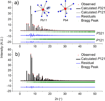
Figure 1. High-resolution synchrotron x-ray diffraction on (a) Pb3TeCo3V2O14 and (b) Pb3TeCo3P2O14. For Pb3TeCo3V2O14, the unit cell was refined using both P321 (black) and P121 (grey). A list of atomic coordinates for the new cell can be found in [21]; (inset) Pb11–O8 polyhedra are significantly distorted as compared to other Pb–O8 polyhedra like Pb4–O8, showing the possibility of lone-pair driven distortion.
Download figure:
Standard image High-resolution imageKrizan et al reason that Pb2+ lone pairs are responsible for the structural distortion causing the supercell. Previous Rietveld analysis on single crystal Pb3TeZn3As2O14 shows a unit cell with P321 space group and only slight asymmetry on PbO8 polyhedra suggesting no lone-pair effect [18]. Fourier difference maps also show no evidence of effective lone pairs. It is worth mentioning that this study took place nearly 15 years ago; diffraction technology has since vastly improved and the supercell reflections found in this study are rather weak. However, in Pb3TeCo3V2O14 and Pb3TeCo3P2O14, there is a wide variety in the shape of PbO8 polyhedra. In particular, the Pb(11)O8 polyhedra are extremely distorted suggesting that, at least in these compounds, Pb2+ lone pairs may play a role in the structural distortion (inset figure 1). These compounds are not the only Ca3Ga4Ge2O14 group members which break trigonal symmetry: Pb3TeZn3P2O14, Pb3TeZn3V2O14, and La3SbZn3Ge2O14 all have superlattice reflections although the true superstructure of La3SbZn3Ge2O14 is still unknown [5]. The latter is particularly interesting as lone pairs do not exist and obviously cannot account for the loss of symmetry. While further study on Pb3TeZn3As2O14 is warranted to determine whether or not a supercell exists, the body of evidence suggests that Pb2+ lone pairs may not be the only force driving the loss of symmetry. The supercell also has important consequences for the magnetism in the dugganites. Co2+ trimers, which are equilateral in the trigonal subcell, are isosceles in the monoclinic cell (figure 2). Six distinct trimers with various Co–Co distances are listed in table 1 for both Pb3TeCo3V2O14 and Pb3TeCo3P2O14. For each trimer, this implies two distinct intratrimer superexchange pathways (totalling at least 12 exchange interactions disregarding intertrimer interactions).
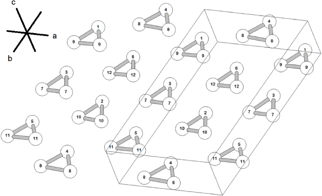
Figure 2. The monoclinic P121 cell alters the equilateral trimer of the P321 subcell into 6 isosceles trimers per unit cell. The unit cell has been shifted down by k = (0,1/2,0) for clarity. Note that trimers with Co atoms 2, 4, and 6 occur in the middle of the unit cell while trimers with Co atoms 1, 3, and 5 occur on the unit cell face. This has important symmetry consequences for the orientation of the moments in the magnetic structures.
Download figure:
Standard image High-resolution imageTable 1. Trimer legend and Co–Co bond distances. See figure 2 for Co labels.
| Name | Centring | Bonds | Co–Co bond distance (Å)Pb3TeCo3V2O14 | Co–Co bond distance (Å)Pb3TeCo3P2O14 | Multiplicity |
|---|---|---|---|---|---|
| 1 | a = 0, c ≈ 1/2 | Co1–Co9 | 3.49(3) | 3.58(4) | 2 |
| Co9–Co9 | 3.71(3) | 3.62(3) | 1 | ||
| 2 | a = 1/2,c ≈ 1/2 | Co2–Co10 | 3.56(4) | 3.76(4) | 2 |
| Co10–Co10 | 3.16(3) | 3.49(3) | 1 | ||
| 3 | a = 0, c ≈ 1/2 | Co3–Co7 | 3.68(4) | 3.59(4) | 2 |
| Co7–Co7 | 3.37(3) | 3.25(3) | 1 | ||
| 4 | a = 1/2,c = 1/2 | Co4–Co8 | 3.71(3) | 3.71(4) | 2 |
| Co8–Co8 | 4.10(3) | 3.76(3) | 1 | ||
| 5 | a = 0, c = 1/2 | Co5–Co11 | 3.45(3) | 3.53(4) | 2 |
| Co11–Co11 | 4.00(3) | 3.36(3) | 1 | ||
| 6 | a = 1/2,c = 1/2 | Co6–Co12 | 3.55(4) | 3.61(3) | 2 |
| Co12–Co12 | 3.43(3) | 3.91(3) | 1 |
Following our refinement of Pb3TeCo3V2O14 to the new supercell, we were prompted to revisit our earlier magnetic structure solutions [20]. Pb3TeCo3V2O14 has two magnetic phase transitions at TN1 = 8.6 K and TN2 = 6.0 K. For all magnetic structural solutions, the SARAh package was used to find the symmetry allowed basis vectors and irreducible representations. TN1 was previously reported to have a propagation vector of k = (0.752,0,1/2). However, while the refinement converged on the most intense peaks of the magnetic phase, smaller peaks were missed. In addition to indexing all of the peaks from the previous study, we were able to index most of the missed peaks using a propagation vector of k = (1/2,0, − 1/2), which was the same one used in the recent magnetic structure refinement of Pb3TeCo3P2O14 [21]. The data were refined according to the irreducible representation (IR) Γ(1) of the little group Gx formed from the space group P121 and the propagation vector, restricted to the Hilbert space defined by basis vectors Ψ(1),Ψ(2), and Ψ(3) [25–28]. Co2+ moments occupying the two-fold symmetrical 1d site (trimers 2, 4, 6) were constrained to point along the b axis in accordance with Γ(1). It was found that a small net moment pointing along the b axis for these trimers significantly improved our refinement. Furthermore, these net moments appear to align in ferromagnetic fashion with respect to each other in each row parallel to b. A c-axis moment component consistent with our symmetry analysis was added to Co occupying the 2e site in these trimers to further improve the fit. For trimers 1, 3, 5, the two-fold symmetrical 1b site Co moments are constrained to point parallel to the ac plane in accordance with Γ(1) while 2e Co moments were left unconstrained leading to net moments on these trimers as well. The results of the refinement are shown in figure 3(a) while figure 3(b) shows the orientation of the moment vectors in real space. It is also noted that the intensity of the magnetic peaks changes quite dramatically in a 2.5 K temperature window (figure 3(c)). From figure 3(b), the moments appear much more disordered than those found in the second magnetic structure.
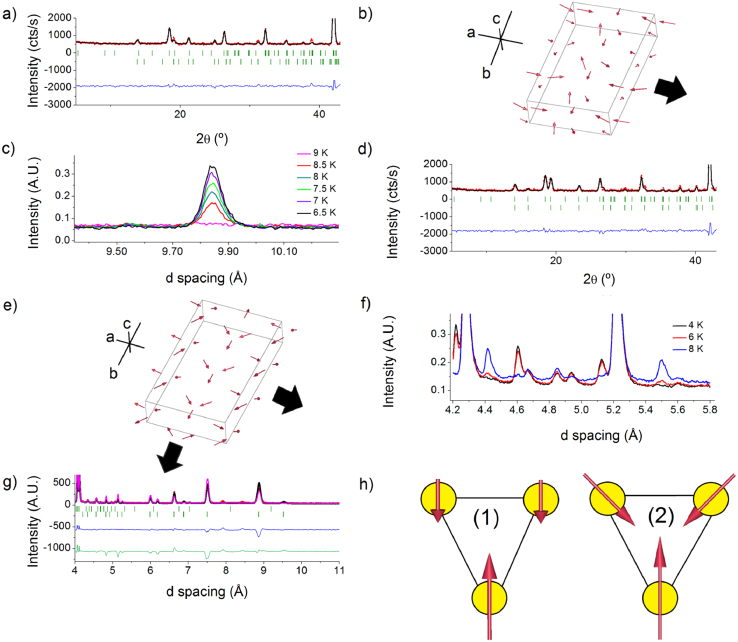
Figure 3. All refinements have the same legend as in figure 1 unless otherwise noted; (a) refinement of the first magnetic structure (TN1) of Pb3TeCo3V2O14; (b) real space orientation of the moments in TN1. Thick arrow denotes direction of the propagation vector (c-axis component not shown); (c) the intensity of all magnetic peaks in TN1 more than doubles over 2.5 K. The magnetic peak in this figure is from (1/2,0, − 1/2) and (1/2,0,1/2) reflections; (d) refinement of the second magnetic structure (TN2) of Pb3TeCo3V2O14; (e) real space orientation of the moments in TN2. Thick arrows denotes direction of the propagation vector (c-axis component not shown); (f) evidence that both magnetic phases coexist at 6.0 K after more than 2 h of thermal equilibration at this temperature; (g) refinement of the magnetic structure of Pb3TeCo3P2O14. The pink curve denotes the fit of the model proposed by Krizan et al [21] while the green line (lower) is the residual to that fit; (h) trimer 4 as depicted by our model (1) and Krizan et al model (2). Note that our model has a small net moment oriented along the two-fold axis.
Download figure:
Standard image High-resolution imageFor TN2, the magnetic structure was discussed in detail previously [20]. As with TN1, the original refinement converged on most peaks, but smaller peaks were missed. We were able to refine this structure using a new propagation vector of k = (1/2,1/2, − 1/2) instead of k = (5/6,5/6,1/2), resolving a prominent issue concerning how the propagation vector could change so abruptly at low temperatures. The data was refined according to IR Γ(1) in much the same manner as TN1, the results of which can be seen in figure 3(d). Figure 3(e) shows the real space orientation of the moments; remarkable agreement is observed between the original magnetic refinement and the new refinement. In the original magnetic structure, a sine wave component on the moment magnitude was observed over 6 trimers in the a direction of the subcell [20]. The origin of this feature is obvious in the new structure: with three Co trimers per row per unit cell and a propagation vector along the diagonal, one obtains commensurate ordering over 6 trimers. For trimers 2, 4, and 6, a much larger net moment along the b axis than that in the first structure led to a stable refinement. Additionally, moments for these trimers were constrained to remain in the ab plane. Moments in trimers 1, 3 and 5 are canted with respect to the c axis. All Co2+ moments ranged between 2.3 and 3.3 μB in this structure, which is short, but within reasonable agreement of the effective moment found from the magnetic susceptibility [20]. Figure 3(f) depicts data from WISH at the ISIS spallation source. In particular, magnetic phase coexistence is observed at 6 K. While this could be due to thermal gradients within the sample, it should be noted that for this run, the sample was equilibrated at 6 K from 8.5 K for over 2 h prior to data collection due to the refilling of the source moderator. This is also in agreement with a sudden drop in the magnetic peak intensity of the first magnetic structure at 6.25 K (not shown), indicating a transition temperature range of at least 0.25 K.
For Pb3TeCo3P2O14, data was taken using WISH at ISIS: a low-background time-of-flight diffractometer. In addition to significant diffuse scattering observed as the transition temperature was approached, similar to that observed earlier for Pb3TeCo3V2O14 [20], one magnetic transition was observed at 12.5 K. The magnetic structure was solved in a similar manner as discussed previously. However, Krizan et al have also reported solving the magnetic structure of this compound by constraining the trimers 2, 4, and 6 to have zero net moment while constraining moments in trimers 1, 3, and 5 to point along the c axis [21]. We compare the results of our magnetic structure model against theirs in figure 3(g). The difference between the models lays in the orientation of Co in the 2e site on trimers 2, 4, and 6: in our model, these Co2+ spins have no moment component along the a axis and were allowed to vary (figure 3(h)). This creates net moments on those trimers that are oriented along the b axis. We also note the presence of two additional peaks that do not belong to either the nuclear or magnetic phases at d = 7.9 Å (Q = 0.79 Å−1) and d = 8.4 Å (Q = 0.74 Å−1). These peaks do not disappear above TN suggesting that they are impurities, however they were not observed in the x-ray data. Upon closer examination, they may appear in the data shown by Krizan et al as well, although our data has a much higher signal-to-noise ratio, allowing us to clearly resolve these peaks. This may suggest that there is an even larger supercell for this dugganite and would be consistent with the anomalous rattling of P–O tetrahedra reported by Krizan et al as well as the peak absence from the x-ray data. Even if a larger supercell is present, we show that the magnetism can be well modelled with the current cell.
The investigation of the magnetic structures of the Co–V and Co–P containing dugganites prompted us to study what happens as P is doped onto the V site (figure 4(a)). Pb3TeCo3V2−xPxO14 (x = 0.5,x = 1.0) samples were prepared by conventional solid state reaction. While the possibility of two distinct phases cannot be ruled out without high-resolution synchrotron data, clues can be drawn suggesting the contrary. For example, the colours of the doped samples are intermediate to that of Pb3TeCo3V2O14 (slate blue) and Pb3TeCo3P2O14 (brilliant indigo). Also, the lattice constants match that for doping according to Vegard's law (figure 4(a)) [29]. A small anomaly is noted when using the c parameter for Vegard's law (not shown) and likely reflects slightly larger-than-reported standard deviations of refined parameters. The structure was refined using a conventional laboratory x-ray diffractometer using the same supercell as the other dugganites (figure 4(b)). As a simplification, only the instrumental parameters, lattice constants, and site occupancies were refined. The V–P site occupancies were refined equally for all unique V sites. V–O and P–O bond lengths differ considerably, and as a result, this is not the best approximation to make. However, the lack of high-resolution x-ray and neutron data would not allow us to satisfactorily characterize the cationic or O sublattices of the doped dugganites any further than this. It is again likely that the actual unit cell is an even larger supercell than the one used [21]. The final clue hinting at a single phase is that the samples have only one magnetic transition at temperatures intermediate to those of the pure V and pure P containing dugganites (figure 4(c)). Like the end series members, significant diffuse scattering is observed in both samples, but particularly at temperatures as low as 7 K for the x = 0.5 sample, implying either short-range magnetic order, or fluctuating moments leaking into the elastic channel due to a lack of resolution (figure 4(d)). Unfortunately, this made refining the magnetic structure, or even obtaining a propagation vector rather difficult. A reverse Monte Carlo (RMC) algorithm in SARAh refine (40 RMC cycles, 7 FullProf least squares cycles) was used to find a propagation vector. An exhaustive map of symmetry allowed propagation vectors is presented in figures 4(e) and (f). Each vector was tested starting from those with the lowest χ2 value. While the magnetic structure could not be solved, this does not mean that important conclusions cannot be drawn from the data. Firstly, the propagation vectors for both the x = 0.5 and 1.0 compounds is notably different from either magnetic structure of Pb3TeCo3V2O14 and from Pb3TeCo3P2O14 despite weak chemical pressure/nonmagnetic site doping. Assuming completely random site V/P site-mixing, this implies that weak fluctuations in the internal magnetic field of Co2+ can seriously perturb the magnetism (i.e. the magnetic states in these Co containing dugganites are very delicate). We demonstrate this with measurements of the linear susceptibility of the end series members under weak fields.
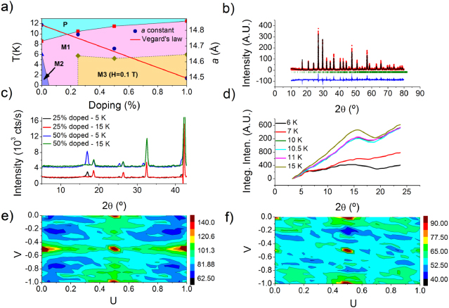
Figure 4. (a) Low-temperature magnetic phase diagram as a function of doping showing the paramagnetic (P), first antiferromagnetic ordered (M1), second antiferromagnetic ordered (M2), and field-induced ordered (M3) states. Note that where the M2 region ends as a function of doping is currently not known; (b) refinement of Pb3TeCo3V1.5P0.5O14 to the P121 supercell. A low signal-to-noise ratio has prevented us from performing an in-depth crystallographic analysis. This is an avenue for further study; (c) magnetic phases of Pb3TeCo3V2−xPxO14 (x = 0.5,1) using neutron scattering. Both samples exhibit significant diffuse scattering, as well as the same (or similar) propagation vectors; (d) integrated diffuse scattering as a function of temperature for Pb3TeCo3V1.5P0.5O14. The data was smoothed using the Savitzky–Golay function (10-point window) before integrating. Nuclear and magnetic Bragg peaks were also removed before the analysis. 5 K (base) data was subtracted from each temperature; exhaustive list of symmetry allowed propagation vectors (e) k = (U,0,V) and (f) k = (U,1/2,V) were manually tested on the doped magnetic structures from lowest χ2 (blue) to highest χ2 (maroon). Propagation vectors were first run through SARAh refine to determine χ2 values. Although we were unable to solve the structure at this time, we note that the high symmetry propagation vectors k = (1/2,0, − 1/2) and k = (1/2,1/2, − 1/2) used for the magnetic structures of undoped and fully doped series members are actually the worst fits to the data.
Download figure:
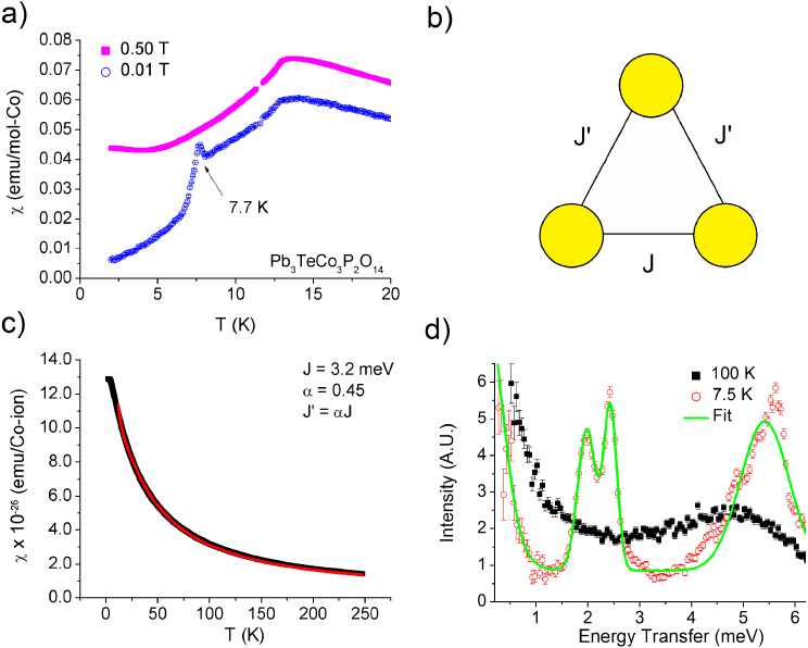
Standard image High-resolution imageAn investigation of the DC-magnetic susceptibility for the dugganite series reveals broad maxima at low temperatures as previously reported for Pb3TeCo3V2O14, followed by sharper features [20]. The broad maxima have been correlated through previous measurements to the appearance of diffuse features in the neutron diffraction profile and are ascribed to the short-ranged ordering of trimer clusters, while the sharper features have been attributed to magnetic ordering. For Pb3TeCo3P2O14, a second zero-field magnetic phase transition is absent, although one can be induced in fields as low as 0.01 T (figure 5(a)). Remarkably, this feature broadens as the field is raised to 0.05 T (not shown) and completely vanishes by 0.5 T, which explains why this transition was missed in earlier studies [21]. It is also worth noting that there is a finite susceptibility as the base temperature of 2 K is reached. This is due to the trimer nature of the system, which prohibits a cancellation of the moment as might be expected for a dimer or tetramer system. The low-temperature magnetic structures across the series confirm that there is always a net ferromagnetic moment in each magnetic unit cell as well.
Figure 5. (a) DC-magnetic susceptibility of Pb3TeCo3P2O14. A low-T second magnetic phase can be induced in fields as low as 0.01 T in this sample; (b) the effective S = 1/2 isosceles trimer model used to fit the susceptibility data [30] where J' = αJ; (c) the magnetic susceptibility of Pb3TeCo3V2O14 is well described by isosceles trimer model (red) [30] using an exchange energy of J = 3.2(1) meV and α = 0.45. A field of 9 T was used in the measurement; (d) Inelastic neutron scattering data taken at SEQUOIA at the SNS using an incident neutron energy of Ei = 8 meV (resolution is approximately 2.1% of the incident energy). The peaks were fitted to a Gaussian function over an exponential background. The broadness in these features likely reflects the different values of J due to the uniqueness of each trimer in the unit cell. Therefore, calculated J and J' energies should be taken as averages.
Download figure:
Standard image High-resolution image
With the detailed structure of the dugganites series now known, we can perform a more quantitative analysis of the susceptibility data. Using a modified form of the effective S = 1/2 isosceles trimer model detailed by Haraldsen, Barnes, and Musfeldt [30] (figure 5(b)), the DC-magnetic susceptibility above the magnetic transitions in Pb3TeCo3V2O14 was fit to:
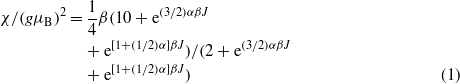
where J corresponds to the base spin–spin exchange constant, and J = αJ is the exchange constant for the identical bond lengths of the trimer (as outlined in figures 5(b), (c) and defined in Haraldsen et al [30]). The fits to the susceptibility yield J = 3.15(2) meV and α = 0.45(1). These parameters can be directly compared to the inelastic neutron scattering data taken using the SEQUOIA spectrometer at the SNS on Pb3TeCo3V2O14 with a standard ILL cryostat. At high temperatures, broad dispersionless features were noted in the excitation spectrum that sharpen as the temperature is lowered. Below TN1, three prominent excitations are noted, which were fitted to Gaussian peaks over an exponential background (figure 5(d)). Since there are no prominent spin waves, and only weak coupling between the trimers, we ascribe these three transitions to the first three energy levels of the isosceles trimer—a split degenerate ground state energy level, and the first excited state [30]. Using the J and α parameters from the susceptibility, the first three allowed neutron scattering transitions allowing for the selection rule of ΔS = 0, ± 1 should occur at ΔE1 = 1.6(1) meV,ΔE2 = 2.4(1) meV and ΔE3 = 5.2(1) meV. Our fit yielded excitations at ΔE1 = 1.978(5) meV,ΔE2 = 2.432(4) meV and ΔE3 = 5.41(4) meV although considerable broadness is observed for ΔE3. This is likely an artefact resulting from averaging all exchange energies into two terms. Nevertheless, the isolated isosceles trimer model represents the data quite well. Improvements to the model based on the exchange pathways and distribution of trimer sizes would likely result in a more precise fit to the data and account for the broadness in the features (due to different trimer bond lengths).
4. Conclusion
In this study, we have provided a comprehensive study of the magnetic properties of the dugganite series Pb3TeCo3A2O14 (A = V, P). With the larger structural unit cell, we have completed an analysis of the magnetic structures, and proposed a weakly interacting effective S = 1/2 isosceles trimer model to explain the magnetic susceptibility and the inelastic neutron scattering results. For the first time, the elucidation of these properties, coupled with the neutron data, enables the construction of a phase diagram for the doped series. While the potential for multiferroic properties has only been tested for the parent compound Pb3TeCo3V2O14, the magnetic phase diagram as a function of doping is essential for other tests across the series and to guide theorists for future investigation of other dugganite compounds, such as Ba3TeCo3P2O14 [21]. We conclude by noting that the rich magnetic phase diagram for the different members of this series also provides an opportunity to study other exotic magnetic phases, as well as the possibility of a multiferroic quantum critical point in high applied fields [31].
Acknowledgments
We would like to thank NSERC, The ACS Petroleum Fund, and the CFI for funding this project. In addition, HJS graciously thanks the Vanier CGS (NSERC), MGS, and the University of Manitoba for funding. KC-K would like to acknowledge the NSERC USRA program for funding. CRW thanks the Canada Research Chair (Tier II) program for additional funding. We would also like to thank the support staff at the Canadian Neutron Beam Centre in Chalk River, as well as the staff at the Advanced Photon Source at Argonne, IL. Portions of this research at the Oak Ridge National Laboratory's SNS and Argonne National Laboratory's APS were sponsored by the Scientific User Facilities Division, Office of Basic Energy Sciences, US Department of Energy (APS under Contract No. DE-AC02-06CH11357). Experiments at the ISIS Pulsed Neutron Source were supported by a beamtime allocation from the Science and Technology Facilities Council. The authors would like to thank T Sherline, D Khalyavin and M Bieringer for useful discussions.